Cn|En

现货抗体产品库

LC3A(5G10)Mouse Monoclonal Antibody

产品详情 相关文献 产品问答 相关产品

产品基本信息

产品货号
BD-PM3649
产品名称
LC3A(5G10)Mouse Monoclonal Antibody
别名
Microtubule-associated proteins 1A/1B light chain 3A (Autophagy-related protein LC3 A) (Autophagy-related ubiquitin-like modifier LC3 A) (MAP1 light chain 3-like protein 1) (MAP1A/MAP1B light chain 3 A) (MAP1A/MAP1B LC3 A) (Microtubule-associated protein 1 light chain 3 alpha)
类别
常规抗体
基因名称
MAP1LC3A
蛋白名称
LC3A
推荐应用
WB
反应种属
Human,Rat,Mouse
浓度
1 mg/ml
存储缓冲液
Liquid in PBS containing 50% glycerol, 0.5% BSA and 0.02% New type preservative N.
Human Gene ID
84557
Human Gene Link
http://www.ncbi.nlm.nih.gov/sites/entrez?db=gene&term=84557
Human Swissprot No.
Q9H492
Human Swissprot Link
http://www.uniprot.org/uniprotkb/Q9H492/entry
Mouse Swissprot No.
Q91VR7
Mouse Swissprot Link
http://www.uniprot.org/uniprot/Q91VR7
Rat Swissprot No.
Q6XVN8
Rat Swissprot Link
http://www.uniprot.org/uniprot/O54941Q6XVN8
免疫原
Synthetic Peptide of LC3A
特异性
LC3A protein detects endogenous levels of LC3A
稀释度
WB 1:1000-2000, IHC 1:100-200 IF 1:200
预测分子量
14,16kD
运输及保存条件
-20°C/1 year
宿主
Monoclonal, Mouse
背景介绍
MAP1A and MAP1B are microtubule-associated proteins which mediate the physical interactions between microtubules and components of the cytoskeleton. MAP1A and MAP1B each consist of a heavy chain subunit and multiple light chain subunits. The protein encoded by this gene is one of the light chain subunits and can associate with either MAP1A or MAP1B. Two transcript variants encoding different isoforms have been found for this gene. The expression of variant 1 is suppressed in many tumor cell lines, suggesting that may be involved in carcinogenesis. [provided by RefSeq, Feb 2012],
组织表达
Most abundant in heart, brain, liver, skeletal muscle and testis but absent in thymus and peripheral blood leukocytes.
细胞定位
Cytoplasmic vesicle, autophagosome membrane ; Lipid-anchor . Endomembrane system ; Lipid-anchor . Cytoplasm, cytoskeleton . LC3-II binds to the autophagic membranes. .
功能
function:Probably involved in formation of autophagosomal vacuoles (autophagosomes).,PTM:The precursor molecule is cleaved by APG4B/ATG4B to form the cytosolic form, LC3-I. This is activated by APG7L/ATG7, transferred to ATG3 and conjugated to phospholipid to form the membrane-bound form, LC3-II.,similarity:Belongs to the MAP1 LC3 family.,subcellular location:LC3-II binds to the autophagic membranes.,subunit:3 different light chains, LC1, LC2 and LC3, can associate with MAP1A and MAP1B proteins.,tissue specificity:Most abundant in heart, brain, liver, skeletal muscle and testis but absent in thymus and peripheral blood leukocytes.,
期货
现货
纯化
The antibody was affinity-purified from mouse ascites by affinity-chromatography using specific immunogen.

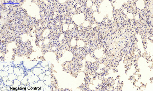
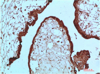
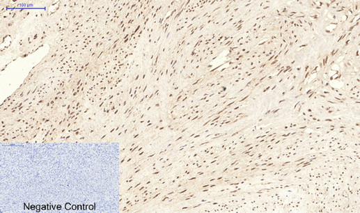
Immunohistochemical analysis of paraffin-embedded Human-uterus tissue. 1,LC3A Mouse Monoclonal Antibody(5G10) was diluted at 1:200(4°C,overnight). 2, Sodium citrate pH 6.0 was used for antibody retrieval(>98°C,20min). 3,Secondary antibody was diluted at 1:200(room tempeRature, 30min). Negative control was used by secondary antibody only.

Immunohistochemical analysis of paraffin-embedded Rat-kidney tissue. 1,LC3A Mouse Monoclonal Antibody(5G10) was diluted at 1:200(4°C,overnight). 2, Sodium citrate pH 6.0 was used for antibody retrieval(>98°C,20min). 3,Secondary antibody was diluted at 1:200(room tempeRature, 30min). Negative control was used by secondary antibody only.

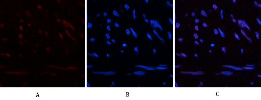
Immunofluorescence analysis of Human-lung-cancer tissue. 1,LC3A Mouse Monoclonal Antibody(5G10)(red) was diluted at 1:200(4°C,overnight). 2, Cy3 labled Secondary antibody was diluted at 1:300(room temperature, 50min).3, Picture B: DAPI(blue) 10min. Picture A:Target. Picture B: DAPI. Picture C: merge of A+B
Immunofluorescence analysis of Mouse-spleen tissue. 1,LC3A Mouse Monoclonal Antibody(5G10)(red) was diluted at 1:200(4°C,overnight). 2, Cy3 labled Secondary antibody was diluted at 1:300(room temperature, 50min).3, Picture B: DAPI(blue) 10min. Picture A:Target. Picture B: DAPI. Picture C: merge of A+B

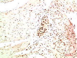
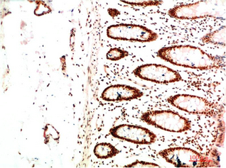
Immunohistochemical analysis of paraffin-embedded Human Heptacarcinoma Tissue using LC3A Mouse mAb diluted at 1:200.

Immunohistochemical analysis of paraffin-embedded Human Prostate Carcinoma Tissue using LC3A Mouse mAb diluted at 1:200.

Western blot analysis of 1) Hela Cell Lysate, 2) 3T3 Cell Lysate, 3) Rat Brain Tissue Lysate using LC3A Mouse mAb diluted at 1:1000.

相关文献

产品问答

相关产品

免责声明| 法律支持| 联系方式

市场:027-65023363   行政/人事:027-62439686   邮箱:marketing@brainvta.com  

销售总监:张经理  18995532642  华东区:陈经理 18013970337   华南区:王经理 13100653525   华中/西区:杨经理 18186518905   华北区:张经理 18893721749

地址:中国武汉东湖高新区光谷七路128号中科开物产业园1号楼

Copyright © 武汉枢密脑科学技术有限公司. All RIGHTS RESERVED.
鄂ICP备2021009124号 DIGITAL BY VTHINK