产品名称
Smad3(4C9)Mouse Monoclonal Antibody
存储缓冲液
Liquid in PBS containing 50% glycerol, 0.5% BSA and 0.02% New type preservative N.
Human Gene Link
http://www.ncbi.nlm.nih.gov/sites/entrez?db=gene&term=4088
Human Swissprot No.
P84022
Human Swissprot Link
http://www.uniprot.org/uniprotkb/P84022/entry
Mouse Swissprot No.
Q8BUN5
Mouse Swissprot Link
http://www.uniprot.org/uniprot/Q8BUN5
Rat Swissprot Link
http://www.uniprot.org/uniprot/O54941P84025
免疫原
Synthetic Peptide of Smad3 at AA range of 350-430
特异性
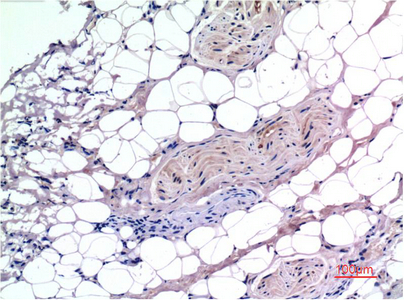
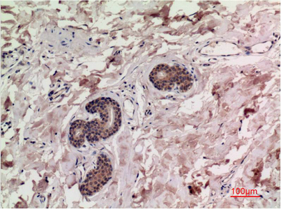
Smad3 protein detects endogenous levels of SMAD3
背景介绍
The protein encoded by this gene belongs to the SMAD, a family of proteins similar to the gene products of the Drosophila gene 'mothers against decapentaplegic' (Mad) and the C. elegans gene Sma. SMAD proteins are signal transducers and transcriptional modulators that mediate multiple signaling pathways. This protein functions as a transcriptional modulator activated by transforming growth factor-beta and is thought to play a role in the regulation of carcinogenesis. [provided by RefSeq, Apr 2009],
组织表达
Brain,Colon carcinoma,Esophagus tumor,Pancreas,Placenta,Spleen,Umbilical cord blood
细胞定位
Cytoplasm . Nucleus . Cytoplasmic and nuclear in the absence of TGF-beta. On TGF-beta stimulation, migrates to the nucleus when complexed with SMAD4 (PubMed:15799969, PubMed:21145499). Through the action of the phosphatase PPM1A, released from the SMAD2/SMAD4 complex, and exported out of the nucleus by interaction with RANBP1 (PubMed:16751101, PubMed:19289081). Co-localizes with LEMD3 at the nucleus inner membrane (PubMed:15601644). MAPK-mediated phosphorylation appears to have no effect on nuclear import (PubMed:19218245). PDPK1 prevents its nuclear translocation in response to TGF-beta (PubMed:17327236). Localized mainly to the nucleus in the early stages of embryo development with expression becoming evident in the cytoplasm of the inner cell mass at the blastocyst stage (By similarity). .
信号通路
Cell_Cycle_G1S;Cell_Cycle_G2M_DNA;WNT;WNT-T CELLTGF-beta;Adherens_Junction;Pathways in cancer;Colorectal cancer;Pancreatic cancer;Chronic myeloid leukemia;
功能
disease:Defects in SMAD3 may be a cause of colorectal cancer (CRC) [MIM:114500].,domain:The MH2 domain is sufficient to carry protein nuclear export.,function:Transcriptional modulator activated by TGF-beta (transforming growth factor) and activin type 1 receptor kinase. SMAD3 is a receptor-regulated SMAD (R-SMAD).,PTM:Phosphorylated on serine by TGF-beta and activin type 1 receptor kinases.,similarity:Belongs to the dwarfin/SMAD family.,similarity:Contains 1 MH1 (MAD homology 1) domain.,similarity:Contains 1 MH2 (MAD homology 2) domain.,subcellular location:In the cytoplasm in the absence of ligand. Migration to the nucleus when complexed with Smad4.,subunit:Interacts with HGS. Interacts with NEDD4L in response to TGF-beta. Interacts with TTRAP (By similarity). Interacts with SARA (SMAD anchor for receptor activation); form trimers with another SMAD3 and the co-SMAD SMAD4. Interacts with JUN/FOS, vitamin D receptor, homeobox protein TGIF and TGIF2, PEBP2-alpha C subunit, CREB-binding protein (CBP), p300, SKI, SNON, ATF2, SMURF2, AIP1, DACH1 and TGFB1I1. Part of a complex consisting of AIP1, ACVR2A, ACVR1B and SMAD3. Found in a complex with SMAD2 and TRIM33 upon addition of TGF-beta. Interacts with SMAD2 and TRIM33. Found in a complex with SMAD3, Ran and XPO4. Interacts with XPO4. Interacts with LBXCOR1 and CORL2.,
纯化
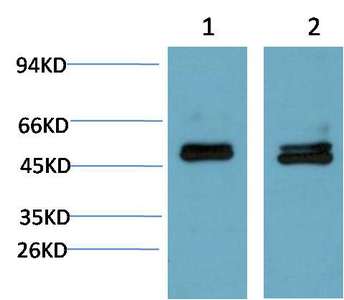
The antibody was affinity-purified from mouse ascites by affinity-chromatography using specific immunogen.