产品名称
Flt3-L Rabbit Polyclonal Antibody
别名
FLT3LG; Fms-related tyrosine kinase 3 ligand; Flt3 ligand; Flt3L; SL cytokine
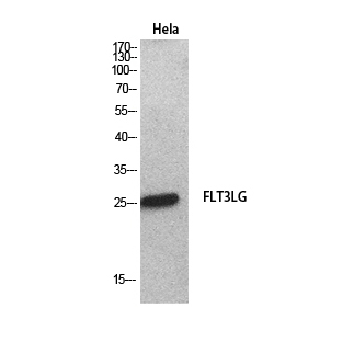
蛋白名称
Fms-related tyrosine kinase 3 ligand
存储缓冲液
Liquid in PBS containing 50% glycerol, 0.5% BSA and 0.02% New type preservative N.
Human Gene Link
http://www.ncbi.nlm.nih.gov/sites/entrez?db=gene&term=2323
Human Swissprot No.
P49771
Human Swissprot Link
http://www.uniprot.org/uniprotkb/P49771/entry
Mouse Swissprot No.
P49772
Mouse Swissprot Link
http://www.uniprot.org/uniprot/P49772
免疫原
The antiserum was produced against synthesized peptide derived from the C-terminal region of human FLT3LG. AA range:171-220
特异性
Flt3-L Polyclonal Antibody detects endogenous levels of Flt3-L protein.
宿主
Polyclonal, Rabbit,IgG
背景介绍
Dendritic cells (DCs) provide the key link between innate and adaptive immunity by recognizing pathogens and priming pathogen-specific immune responses. FLT3LG controls the development of DCs and is particularly important for plasmacytoid DCs and CD8 (see MIM 186910)-positive classical DCs and their CD103 (ITGAE; MIM 604682)-positive tissue counterparts (summary by Sathaliyawala et al., 2010 [PubMed 20933441]).[supplied by OMIM, Jan 2011],
细胞定位
[Isoform 1]: Cell membrane; Single-pass type I membrane protein.; [Isoform 2]: Secreted.
信号通路
Cytokine-cytokine receptor interaction;Hematopoietic cell lineage;Pathways in cancer;
功能
function:Stimulates the proliferation of early hematopoietic cells. Synergizes well with a number of other colony stimulating factors and interleukins.,subunit:Homodimer (isoform 2).,
纯化
The antibody was affinity-purified from rabbit antiserum by affinity-chromatography using epitope-specific immunogen.