产品名称
CD294 Rabbit Polyclonal Antibody
别名
PTGDR2; CRTH2; DL1R; GPR44; Prostaglandin D2 receptor 2; Chemoattractant receptor-homologous molecule expressed on Th2 cells; G-protein coupled receptor 44; CD294
蛋白名称
Prostaglandin D2 receptor 2
存储缓冲液
Liquid in PBS containing 50% glycerol, 0.5% BSA and 0.02% New type preservative N.
Human Gene Link
http://www.ncbi.nlm.nih.gov/sites/entrez?db=gene&term=11251
Human Swissprot No.
Q9Y5Y4
Human Swissprot Link
http://www.uniprot.org/uniprotkb/Q9Y5Y4/entry
Mouse Gene Link
http://www.ncbi.nlm.nih.gov/sites/entrez?db=gene&term=14764
Mouse Swissprot No.
Q9Z2J6
Mouse Swissprot Link
http://www.uniprot.org/uniprot/Q9Z2J6
Rat Gene Link
http://www.ncbi.nlm.nih.gov/sites/entrez?db=gene&term=309212
Rat Swissprot Link
http://www.uniprot.org/uniprot/Q6XKD3
免疫原
The antiserum was produced against synthesized peptide derived from the Internal region of human PTGDR2. AA range:161-210
特异性
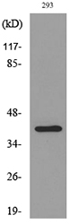
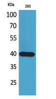
CD294 Polyclonal Antibody detects endogenous levels of CD294 protein.
宿主
Polyclonal, Rabbit,IgG
背景介绍
This gene encodes a G-protein-coupled receptor that is preferentially expressed in CD4+ effector T helper 2 (Th2) cells. This protein is a prostaglandin D2 receptor that mediates the pro-inflammatory chemotaxis of eosinophils, basophils, and Th2 lymphocytes generated during allergic inflammation. Single nucleotide polymorphisms in the 3' UTR of this gene have been associated with asthma susceptibility.[provided by RefSeq, Mar 2011],
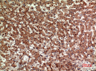
组织表达
Widespread expression. High expression in stomach, small intestine, heart and thymus. Intermediate expression in colon, spinal cord and peripheral blood and low expression in brain, skeletal muscle and spleen. Expressed also on Th2- and Tc2- type cells, eosinophils and basophils.
细胞定位
Cell membrane ; Multi-pass membrane protein . Internalized receptors colocalized with RAB11A. .
功能
function:Orphan receptor.,similarity:Belongs to the G-protein coupled receptor 1 family.,
纯化
The antibody was affinity-purified from rabbit antiserum by affinity-chromatography using epitope-specific immunogen.
.jpg)