产品名称
IGF-IIR Rabbit Polyclonal Antibody
别名
IGF2R; MPRI; Cation-independent mannose-6-phosphate receptor; CI Man-6-P receptor; CI-MPR; M6PR; 300 kDa mannose 6-phosphate receptor; MPR 300;Insulin-like growth factor 2 receptor; Insulin-like growth factor II receptor; IGF-II receptor; M6P/IGF2 receptor; M6P/IGF2R; CD222
蛋白名称
Cation-independent mannose-6-phosphate receptor
存储缓冲液
Liquid in PBS containing 50% glycerol, 0.5% BSA and 0.02% New type preservative N.
Human Gene Link
http://www.ncbi.nlm.nih.gov/sites/entrez?db=gene&term=3482
Human Swissprot No.
P11717
Human Swissprot Link
http://www.uniprot.org/uniprotkb/P11717/entry
Mouse Swissprot No.
Q07113
Mouse Swissprot Link
http://www.uniprot.org/uniprot/Q07113
免疫原
The antiserum was produced against synthesized peptide derived from the C-terminal region of human IGF2R. AA range:2251-2300
特异性
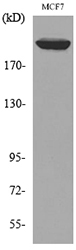
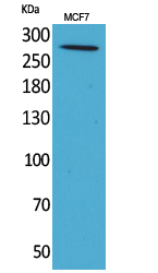
IGF-IIR Polyclonal Antibody detects endogenous levels of IGF-IIR protein.
宿主
Polyclonal, Rabbit,IgG
背景介绍
This gene encodes a receptor for both insulin-like growth factor 2 and mannose 6-phosphate. The binding sites for each ligand are located on different segments of the protein. This receptor has various functions, including in the intracellular trafficking of lysosomal enzymes, the activation of transforming growth factor beta, and the degradation of insulin-like growth factor 2. Mutation or loss of heterozygosity of this gene has been association with risk of hepatocellular carcinoma. The orthologous mouse gene is imprinted and shows exclusive expression from the maternal allele; however, imprinting of the human gene may be polymorphic, as only a minority of individuals showed biased expression from the maternal allele (PMID:8267611). [provided by RefSeq, Nov 2015],
组织表达
Brain,Epithelium,Liver,
细胞定位
Golgi apparatus membrane ; Single-pass type I membrane protein . Endosome membrane ; Single-pass type I membrane protein . Mainly localized in the Golgi at steady state and not detectable in lysosome (PubMed:18817523). Colocalized with DPP4 in internalized cytoplasmic vesicles adjacent to the cell surface (PubMed:10900005). .
功能
domain:Contains 15 repeating units of approximately 147 AA. The most highly conserved region within the repeat consists of a stretch of 13 AA that contains cysteines at both ends.,function:Transport of phosphorylated lysosomal enzymes from the Golgi complex and the cell surface to lysosomes. Lysosomal enzymes bearing phosphomannosyl residues bind specifically to mannose-6-phosphate receptors in the Golgi apparatus and the resulting receptor-ligand complex is transported to an acidic prelyosomal compartment where the low pH mediates the dissociation of the complex. This receptor also binds IGF2.,similarity:Belongs to the MRL1/IGF2R family.,similarity:Contains 1 fibronectin type-II domain.,subunit:Binds GGA1, GGA2 and GGA3.,
纯化
The antibody was affinity-purified from rabbit antiserum by affinity-chromatography using epitope-specific immunogen.