产品名称
ADM Rabbit Polyclonal Antibody
存储缓冲液
Liquid in PBS containing 50% glycerol, 0.5% BSA and 0.02% New type preservative N.
Human Gene Link
http://www.ncbi.nlm.nih.gov/sites/entrez?db=gene&term=133
Human Swissprot No.
P35318
Human Swissprot Link
http://www.uniprot.org/uniprotkb/P35318/entry
Mouse Swissprot No.
P97297
Mouse Swissprot Link
http://www.uniprot.org/uniprot/P97297
Rat Gene Link
http://www.ncbi.nlm.nih.gov/sites/entrez?db=gene&term=25026
Rat Swissprot Link
http://www.uniprot.org/uniprot/P43145
免疫原
The antiserum was produced against synthesized peptide derived from the Internal region of human ADM. AA range:101-150
特异性
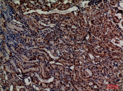
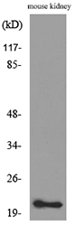
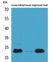
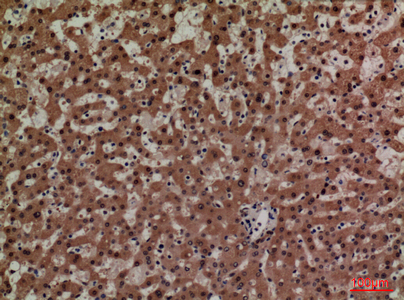
ADM Polyclonal Antibody detects endogenous levels of ADM protein.
宿主
Polyclonal, Rabbit,IgG
背景介绍
The protein encoded by this gene is a preprohormone which is cleaved to form two biologically active peptides, adrenomedullin and proadrenomedullin N-terminal 20 peptide. Adrenomedullin is a 52 aa peptide with several functions, including vasodilation, regulation of hormone secretion, promotion of angiogenesis, and antimicrobial activity. The antimicrobial activity is antibacterial, as the peptide has been shown to kill E. coli and S. aureus at low concentration. [provided by RefSeq, Aug 2014],
组织表达
Highest levels found in pheochromocytoma and adrenal medulla. Also found in lung, ventricle and kidney tissues.
功能
function:AM and PAMP are potent hypotensive and vasodilatator agents. Numerous actions have been reported most related to the physiologic control of fluid and electrolyte homeostasis. In the kidney, am is diuretic and natriuretic, and both am and pamp inhibit aldosterone secretion by direct adrenal actions. In pituitary gland, both peptides at physiologically relevant doses inhibit basal ACTH secretion. Both peptides appear to act in brain and pituitary gland to facilitate the loss of plasma volume, actions which complement their hypotensive effects in blood vessels.,similarity:Belongs to the adrenomedullin family.,tissue specificity:Highest levels found in pheochromocytoma and adrenal medulla. Also found in lung, ventricle and kidney tissues.,
纯化
The antibody was affinity-purified from rabbit antiserum by affinity-chromatography using epitope-specific immunogen.
.jpg)
.jpg)