产品名称
VE-Cadherin Rabbit Polyclonal Antibody
别名
CDH5; Cadherin-5; 7B4 antigen; Vascular endothelial cadherin; VE-cadherin; CD antigen CD144
存储缓冲液
Liquid in PBS containing 50% glycerol, 0.5% BSA and 0.02% New type preservative N.
Human Gene Link
http://www.ncbi.nlm.nih.gov/sites/entrez?db=gene&term=1003
Human Swissprot No.
P33151
Human Swissprot Link
http://www.uniprot.org/uniprotkb/P33151/entry
Mouse Gene Link
http://www.ncbi.nlm.nih.gov/sites/entrez?db=gene&term=12562
Mouse Swissprot No.
P55284
Mouse Swissprot Link
http://www.uniprot.org/uniprot/P55284
免疫原
The antiserum was produced against synthesized peptide derived from human CDH5. AA range:697-746
特异性
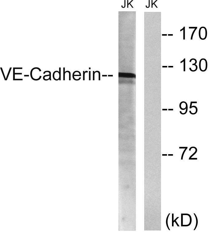
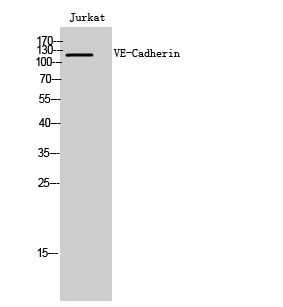
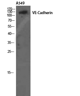
VE-Cadherin Polyclonal Antibody detects endogenous levels of VE-Cadherin protein.
宿主
Polyclonal, Rabbit,IgG
背景介绍
This gene encodes a classical cadherin of the cadherin superfamily. The encoded preproprotein is proteolytically processed to generate the mature glycoprotein. This calcium-dependent cell-cell adhesion molecule is comprised of five extracellular cadherin repeats, a transmembrane region and a highly conserved cytoplasmic tail. Functioning as a classical cadherin by imparting to cells the ability to adhere in a homophilic manner, this protein plays a role in endothelial adherens junction assembly and maintenance. This gene is located in a gene cluster in a region on the long arm of chromosome 16 that is involved in loss of heterozygosity events in breast and prostate cancer. [provided by RefSeq, Nov 2015],
组织表达
Endothelial tissues and brain.
细胞定位
Cell junction . Cell membrane ; Single-pass type I membrane protein . Found at cell-cell boundaries and probably at cell-matrix boundaries. KRIT1 and CDH5 reciprocally regulate their localization to endothelial cell-cell junctions. .
信号通路
Cell adhesion molecules (CAMs);Leukocyte transendothelial migration;
功能
function:Cadherins are calcium dependent cell adhesion proteins.,function:Cadherins are calcium dependent cell adhesion proteins. They preferentially interact with themselves in a homophilic manner in connecting cells; cadherins may thus contribute to the sorting of heterogeneous cell types. This cadherin may play a important role in endothelial cell biology through control of the cohesion and organization of the intercellular junctions. It associates with alpha-catenin forming a link to the cytoskeleton.,similarity:Contains 5 cadherin domains.,subcellular location:Found at cell-cell boundaries and probably at cell-matrix boundaries.,tissue specificity:Endothelial tissues and brain.,
纯化
The antibody was affinity-purified from rabbit antiserum by affinity-chromatography using epitope-specific immunogen.