NSE(13E2)Mouse Monoclonal Antibody
产品基本信息
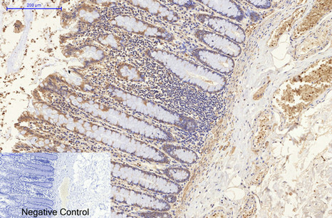
Immunohistochemical analysis of paraffin-embedded Human-colon tissue. 1,NSE Monoclonal Antibody(13E2) was diluted at 1:200(4°C,overnight). 2, Sodium citrate pH 6.0 was used for antibody retrieval(>98°C,20min). 3,Secondary antibody was diluted at 1:200(room tempeRature, 30min). Negative control was used by secondary antibody only.
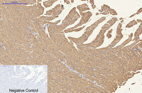
Immunohistochemical analysis of paraffin-embedded Rat-heart tissue. 1,NSE Monoclonal Antibody(13E2) was diluted at 1:200(4°C,overnight). 2, Sodium citrate pH 6.0 was used for antibody retrieval(>98°C,20min). 3,Secondary antibody was diluted at 1:200(room tempeRature, 30min). Negative control was used by secondary antibody only.
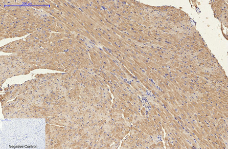
Immunohistochemical analysis of paraffin-embedded Mouse-heart tissue. 1,NSE Monoclonal Antibody(13E2) was diluted at 1:200(4°C,overnight). 2, Sodium citrate pH 6.0 was used for antibody retrieval(>98°C,20min). 3,Secondary antibody was diluted at 1:200(room tempeRature, 30min). Negative control was used by secondary antibody only.
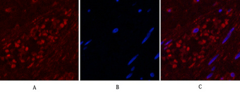
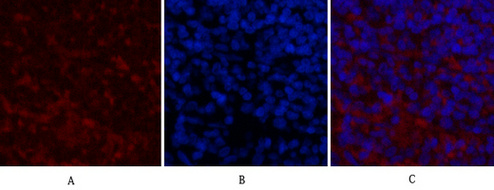
Immunofluorescence analysis of Human-appendix tissue. 1,NSE Monoclonal Antibody(13E2)(red) was diluted at 1:200(4°C,overnight). 2, Cy3 labled Secondary antibody was diluted at 1:300(room temperature, 50min).3, Picture B: DAPI(blue) 10min. Picture A:Target. Picture B: DAPI. Picture C: merge of A+B
Immunofluorescence analysis of Mouse-spleen tissue. 1,NSE Monoclonal Antibody(13E2)(red) was diluted at 1:200(4°C,overnight). 2, Cy3 labled Secondary antibody was diluted at 1:300(room temperature, 50min).3, Picture B: DAPI(blue) 10min. Picture A:Target. Picture B: DAPI. Picture C: merge of A+B
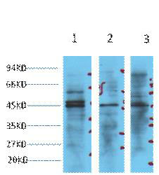
Western blot analysis of 1) Hela, 2) Jurkat, 3) 293T cell lysates, diluted at 1:3000.

IHC staining of Human small cell carcinoma of lung tissue, diluted at 1:200.
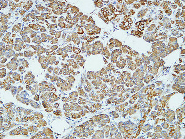
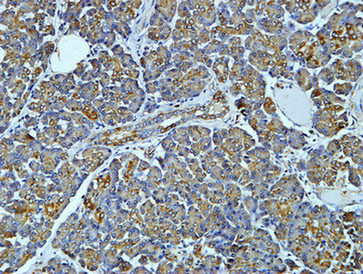
Immunohistochemical analysis of paraffin-embedded Human pancreas. 1, Antibody was diluted at 1:100(4°,overnight). 2, High-pressure and temperature EDTA, pH8.0 was used for antigen retrieval. 3,Secondary antibody was diluted at 1:200(room temperature, 30min).
Immunohistochemical analysis of paraffin-embedded Human pancreas. 1, Antibody was diluted at 1:100(4°,overnight). 2, High-pressure and temperature EDTA, pH8.0 was used for antigen retrieval. 3,Secondary antibody was diluted at 1:200(room temperature, 30min).
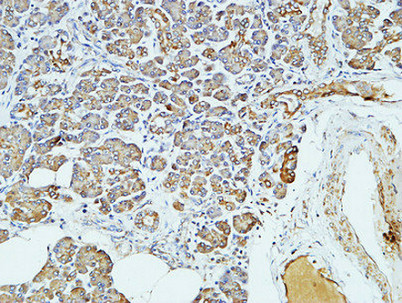
Immunohistochemical analysis of paraffin-embedded Human pancreas. 1, Antibody was diluted at 1:100(4°,overnight). 2, High-pressure and temperature EDTA, pH8.0 was used for antigen retrieval. 3,Secondary antibody was diluted at 1:200(room temperature, 30min).
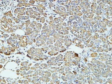
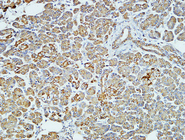
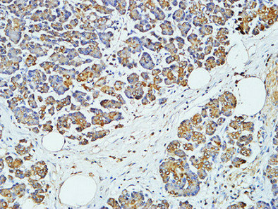
Immunohistochemical analysis of paraffin-embedded Human pancreas. 1, Antibody was diluted at 1:200(4°,overnight). 2, High-pressure and temperature EDTA, pH8.0 was used for antigen retrieval. 3,Secondary antibody was diluted at 1:200(room temperature, 30min).
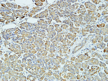
Immunohistochemical analysis of paraffin-embedded Human pancreas. 1, Antibody was diluted at 1:200(4°,overnight). 2, High-pressure and temperature EDTA, pH8.0 was used for antigen retrieval. 3,Secondary antibody was diluted at 1:200(room temperature, 30min).
Immunohistochemical analysis of paraffin-embedded Human pancreas. 1, Antibody was diluted at 1:200(4°,overnight). 2, High-pressure and temperature EDTA, pH8.0 was used for antigen retrieval. 3,Secondary antibody was diluted at 1:200(room temperature, 30min).
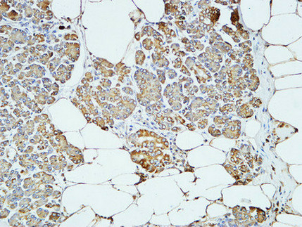
Immunohistochemical analysis of paraffin-embedded Human pancreas. 1, Antibody was diluted at 1:400(4°,overnight). 2, High-pressure and temperature EDTA, pH8.0 was used for antigen retrieval. 3,Secondary antibody was diluted at 1:200(room temperature, 30min).
Immunohistochemical analysis of paraffin-embedded Human pancreas. 1, Antibody was diluted at 1:400(4°,overnight). 2, High-pressure and temperature EDTA, pH8.0 was used for antigen retrieval. 3,Secondary antibody was diluted at 1:200(room temperature, 30min).
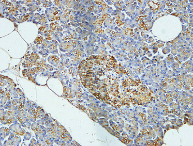
Immunohistochemical analysis of paraffin-embedded Human pancreas. 1, Antibody was diluted at 1:400(4°,overnight). 2, High-pressure and temperature EDTA, pH8.0 was used for antigen retrieval. 3,Secondary antibody was diluted at 1:200(room temperature, 30min).
相关文献
产品问答
相关产品

市场:027-65023363 行政/人事:027-62439686 邮箱:marketing@brainvta.com 客服:18140661572(活动咨询、售后反馈等)
销售总监:张经理 18995532642 华东区:陈经理 18013970337 华南区:王经理 13100653525 华中/西区:杨经理 18186518905 华北区:张经理 18893721749
地址:中国武汉东湖高新区光谷七路128号中科开物产业园1号楼
Copyright © 武汉枢密脑科学技术有限公司. All RIGHTS RESERVED.
鄂ICP备2021009124号 DIGITAL BY VTHINK
