产品名称
HER2(11H9)Mouse Monoclonal Antibody
别名
ERBB2; HER2; MLN19; NEU; NGL; Receptor tyrosine-protein kinase erbB-2; Metastatic lymph node gene 19 protein; MLN 19; Proto-oncogene Neu; Proto-oncogene c-ErbB-2; Tyrosine kinase-type cell surface receptor HER2; p185erbB2; CD340
蛋白名称
Receptor tyrosine-protein kinase erbB-2
存储缓冲液
PBS, pH 7.4, containing 0.5%BSA, 0.02% New type preservative N as Preservative and 50% Glycerol.
Human Gene Link
http://www.ncbi.nlm.nih.gov/sites/entrez?db=gene&term=2064
Human Swissprot No.
P04626
Human Swissprot Link
http://www.uniprot.org/uniprotkb/P04626/entry
Mouse Gene Link
http://www.ncbi.nlm.nih.gov/sites/entrez?db=gene&term=13866
Mouse Swissprot No.
P70424
Mouse Swissprot Link
http://www.uniprot.org/uniprot/P70424
Rat Swissprot Link
http://www.uniprot.org/uniprot/P06494
免疫原
Synthetic Peptide of HER2
特异性
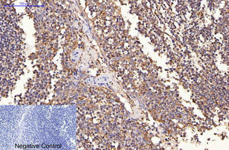
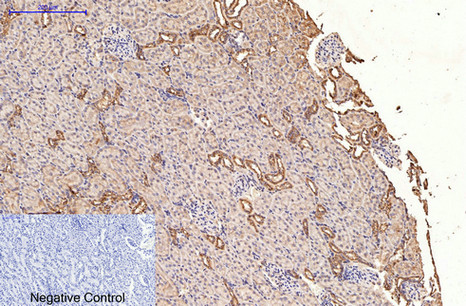
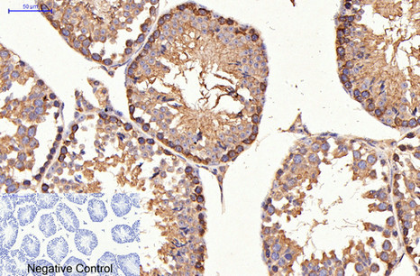
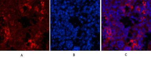
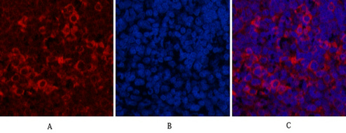
The antibody detects endogenous ErbB-2/HER-2 proteins.
背景介绍
This gene encodes a member of the epidermal growth factor (EGF) receptor family of receptor tyrosine kinases. This protein has no ligand binding domain of its own and therefore cannot bind growth factors. However, it does bind tightly to other ligand-bound EGF receptor family members to form a heterodimer, stabilizing ligand binding and enhancing kinase-mediated activation of downstream signalling pathways, such as those involving mitogen-activated protein kinase and phosphatidylinositol-3 kinase. Allelic variations at amino acid positions 654 and 655 of isoform a (positions 624 and 625 of isoform b) have been reported, with the most common allele, Ile654/Ile655, shown here. Amplification and/or overexpression of this gene has been reported in numerous cancers, including breast and ovarian tumors. Alternative splicing results in several additional transcript variants, some encoding d
组织表达
Expressed in a variety of tumor tissues including primary breast tumors and tumors from small bowel, esophagus, kidney and mouth.
细胞定位
[Isoform 1]: Cell membrane ; Single-pass type I membrane protein. Early endosome . Cytoplasm, perinuclear region. Nucleus. Translocation to the nucleus requires endocytosis, probably endosomal sorting and is mediated by importin beta-1/KPNB1. Also detected in VPS35-positive endosome-to-TGN retrograde vesicles (PubMed:31138794). .; [Isoform 2]: Cytoplasm. Nucleus.; [Isoform 3]: Cytoplasm. Nucleus.
信号通路
ErbB_HER;Calcium;Focal adhesion;Adherens_Junction;Pathways in cancer;Pancreatic cancer;Endometrial cancer;Prostate cancer;Bladder cancer;Non-small cell lung cancer;
功能
catalytic activity:ATP + a [protein]-L-tyrosine = ADP + a [protein]-L-tyrosine phosphate.,disease:Defects in ERBB2 are associated with familial glioma of brain [MIM:137800]; also called glioblastoma multiforme. Gliomas are central nervous system neoplasms derived from glial cells and comprise astrocytomas, glioblastoma multiforme, oligodendrogliomas, and ependymomas.,disease:Defects in ERBB2 are associated with gastric cancer [MIM:137215]; also known as hereditary familial diffuse gastric cancer (HDGC).,disease:Defects in ERBB2 are associated with lung cancer [MIM:211980]; also called adenocarcinoma of lung.,disease:Defects in ERBB2 are associated with ovarian cancer [MIM:167000]. Ovarian cancer is the leading cause of death from gynecologic malignancy. It is characterized by advanced presentation with loco-regional dissemination in the peritoneal cavity and the rare incidence of visceral metastases. These typical features relate to the biology of the disease, which is a principal determinant of outcome.,function:Essential component of a neuregulin-receptor complex, although neuregulins do not interact with it alone. GP30 is a potential ligand for this receptor. Not activated by EGF, TGF-alpha and amphiregulin.,online information:ERBB2 entry,polymorphism:There are fours alleles due to the variations in positions 654 and 655. Allele B1 (Ile-654/Ile-655) has a frequency of 0.782; allele B2 (Ile-654/Val-655) has a frequency of 0.206; allele B3 (Val-654/Val-655) has a frequency of 0.012.,PTM:Ligand-binding increases phosphorylation on tyrosine residues.,similarity:Belongs to the protein kinase superfamily. Tyr protein kinase family. EGF receptor subfamily.,similarity:Contains 1 protein kinase domain.,subunit:Heterodimer with each of the other ERBB receptors (Potential). Interacts with PRKCABP and PLXNB1. Part of a complex with EGFR and either PIK3C2A or PIK3C2B. May interact with PIK3C2B when phosphorylated on Tyr-1196. Interacts with MEMO when phosphorylated on Tyr-1248. Interacts with MUC1. Stimulation by heregulin (HRG) in breast cancer cell lines induces binding of MUC1 with gamma-catenin.,
纯化
The antibody was affinity-purified from mouse ascites by affinity-chromatography using specific immunogen.