产品名称
Translin Rabbit Polyclonal Antibody
别名
TSN; Translin; Component 3 of promoter of RISC; C3PO
存储缓冲液
Liquid in PBS containing 50% glycerol, 0.5% BSA and 0.02% New type preservative N.
Human Gene Link
http://www.ncbi.nlm.nih.gov/sites/entrez?db=gene&term=7247
Human Swissprot No.
Q15631
Human Swissprot Link
http://www.uniprot.org/uniprotkb/Q15631/entry
Mouse Gene Link
http://www.ncbi.nlm.nih.gov/sites/entrez?db=gene&term=22099
Mouse Swissprot No.
Q62348
Mouse Swissprot Link
http://www.uniprot.org/uniprot/Q62348
免疫原
The antiserum was produced against synthesized peptide derived from human TSN. AA range:101-150
特异性
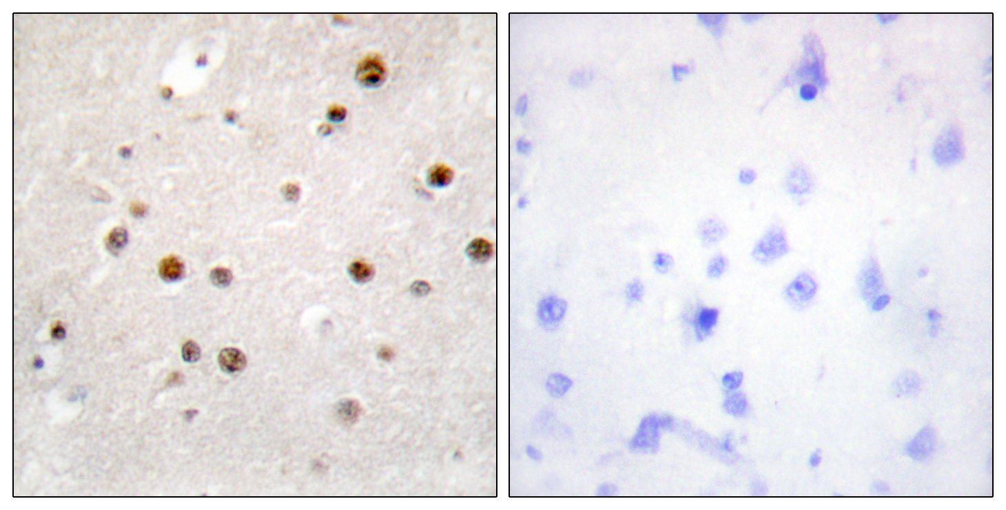
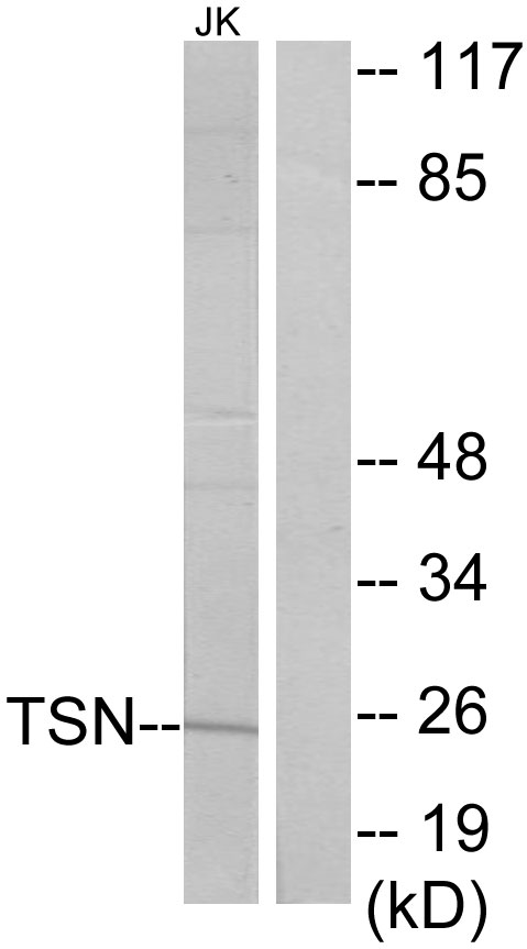
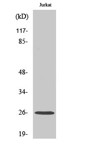
Translin Polyclonal Antibody detects endogenous levels of Translin protein.
宿主
Polyclonal, Rabbit,IgG
背景介绍
This gene encodes a DNA-binding protein which specifically recognizes conserved target sequences at the breakpoint junction of chromosomal translocations. Translin polypeptides form a multimeric structure that is responsible for its DNA-binding activity. Recombination-associated motifs and translin-binding sites are present at recombination hotspots and may serve as indicators of breakpoints in genes which are fused by translocations. These binding activities may play a crucial role in chromosomal translocation in lymphoid neoplasms. This protein encoded by this gene, when complexed with translin-associated protein X, also forms a Mg ion-dependent endoribonuclease that promotes RNA-induced silencing complex (RISC) activation. Alternative splicing results in multiple transcript variants. [provided by RefSeq, May 2012],
细胞定位
Cytoplasm . Nucleus .
功能
function:DNA-binding protein that specifically recognizes consensus sequences at the breakpoint junctions in chromosomal translocations, mostly involving immunoglobulin (Ig)/T-cell receptor gene segments. Seems to recognize single-stranded DNA ends generated by staggered breaks occuring at recombination hot spots.,similarity:Belongs to the translin family.,subunit:Forms a multimeric ring-shaped structure. Interacts with TSNAX.,
纯化
The antibody was affinity-purified from rabbit antiserum by affinity-chromatography using epitope-specific immunogen.