产品名称
THP Rabbit Polyclonal Antibody
别名
UMOD; Uromodulin; Tamm-Horsfall urinary glycoprotein; THP
存储缓冲液
Liquid in PBS containing 50% glycerol, 0.5% BSA and 0.02% New type preservative N.
Human Gene Link
http://www.ncbi.nlm.nih.gov/sites/entrez?db=gene&term=7369
Human Swissprot No.
P07911
Human Swissprot Link
http://www.uniprot.org/uniprotkb/P07911/entry
Mouse Swissprot No.
Q91X17
Mouse Swissprot Link
http://www.uniprot.org/uniprot/Q91X17
免疫原
The antiserum was produced against synthesized peptide derived from human THP. AA range:329-378
特异性
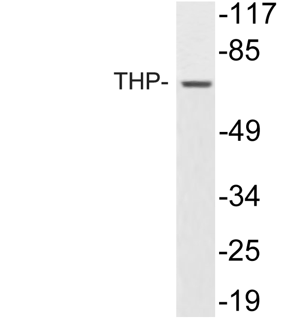
THP Polyclonal Antibody detects endogenous levels of THP protein.
宿主
Polyclonal, Rabbit,IgG
背景介绍
The protein encoded by this gene is the most abundant protein in mammalian urine under physiological conditions. Its excretion in urine follows proteolytic cleavage of the ectodomain of its glycosyl phosphatidylinosital-anchored counterpart that is situated on the luminal cell surface of the loop of Henle. This protein may act as a constitutive inhibitor of calcium crystallization in renal fluids. Excretion of this protein in urine may provide defense against urinary tract infections caused by uropathogenic bacteria. Defects in this gene are associated with the renal disorders medullary cystic kidney disease-2 (MCKD2), glomerulocystic kidney disease with hyperuricemia and isosthenuria (GCKDHI), and familial juvenile hyperuricemic nephropathy (FJHN). Alternative splicing of this gene results in multiple transcript variants. [provided by RefSeq, Jul 2013],
组织表达
Expressed in the tubular cells of the kidney. Most abundant protein in normal urine (at protein level). Synthesized exclusively in the kidney. Expressed exclusively by epithelial cells of the thick ascending limb of Henle's loop (TALH) and of distal convoluted tubule lumen.
细胞定位
Apical cell membrane ; Lipid-anchor, GPI-anchor . Basolateral cell membrane ; Lipid-anchor, GPI-anchor . Cell projection, cilium membrane . Only a small fraction sorts to the basolateral pole of tubular epithelial cells compared to apical localization (PubMed:22776760). Secreted into urine after cleavage (PubMed:18375198, PubMed:26811476). Colocalizes with NPHP1 and KIF3A (PubMed:20172860). .; [Uromodulin, secreted form]: Secreted . Detected in urine. .
功能
disease:Defects in UMOD are a cause of glomerulocystic kidney disease with hyperuricemia and isosthenuria [MIM:609886]. Glomerulocystic kidney disease (GCKD) and medullary cystic disease/familial juvenile hyperuricemic nephropathy (MCKD/HNFJ) are two distinct renal disorders that share some common clinical features. The former is characterized by a cystic dilatation of Bowman's space and a collapse of glomerular tuft. Familial GCKD can be associated with either hypoplastic or normal sized kidneys. A GCKD clinical variant presents the association with hyperuricemia due to low fractional excretion of uric acid and severe impairment of urine concentrating ability that are reminiscent of MCKD/HNFJ.,disease:Defects in UMOD are the cause of familial juvenile hyperuricemic nephropathy (HNFJ) [MIM:162000]. HNFJ is a heritable autosomal dominant renal disease characterized by juvenil onset of hyperuricaemia, polyuria, progressive renal failure, and gout. The disease is associated with interstitial pathological changes resulting in fibrosis.,disease:Defects in UMOD are the cause of medullary cystic kidney disease 2 (MCKD2) [MIM:603860]. MCKD2 and HNFJ constitute a group of heritable renal diseases with a common mode of transmission (autosomal dominant) and shared features including polyuria, hyperuricaemia, progressive renal failure, and gout. Both diseases are associated with interstitial pathological changes resulting in fibrosis. While corticomedullary cysts are well documented in MCKD2, their presence in HNFJ is not well documented. The primary clinical features of MCKD2 and HNFJ vary in presence and severity, complicating the diagnosis of these conditions, particularly in milder cases. Both diseases are considered to be allelic diseases.,function:Not known. May play a role in regulating the circulating activity of cytokines as it binds to IL-1, IL-2 and TNF with high affinity.,similarity:Contains 1 ZP domain.,similarity:Contains 3 EGF-like domains.,subcellular location:Secreted after cleavage in the urine.,tissue specificity:Synthesized by the kidneys and is the most abundant protein in normal human urine.,
纯化
The antibody was affinity-purified from rabbit antiserum by affinity-chromatography using epitope-specific immunogen.