Cn|En

现货抗体产品库

TFDP1 Rabbit Polyclonal Antibody

产品详情 相关文献 产品问答 相关产品

产品基本信息

产品货号
BD-PT4611
产品名称
TFDP1 Rabbit Polyclonal Antibody
别名
TFDP1; DP1; Transcription factor Dp-1; DRTF1-polypeptide 1; DRTF1; E2F dimerization partner 1
类别
常规抗体
基因名称
TFDP1
蛋白名称
Transcription factor Dp-1
推荐应用
WB
反应种属
Human,Rat,Mouse
浓度
1 mg/ml
存储缓冲液
Liquid in PBS containing 50% glycerol, 0.5% BSA and 0.02% New type preservative N.
Human Gene ID
7027
Human Gene Link
http://www.ncbi.nlm.nih.gov/sites/entrez?db=gene&term=7027
Human Swissprot No.
Q14186
Human Swissprot Link
http://www.uniprot.org/uniprotkb/Q14186/entry
Mouse Swissprot No.
Q08639
Mouse Swissprot Link
http://www.uniprot.org/uniprot/Q08639
免疫原
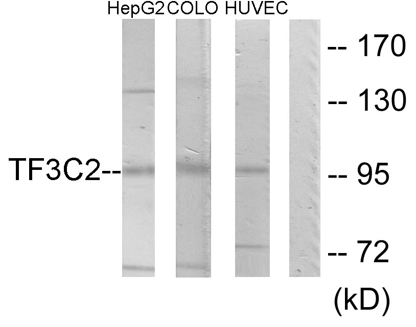
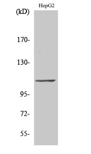
The antiserum was produced against synthesized peptide derived from human DP-1. AA range:361-410
特异性
TFDP1 Polyclonal Antibody detects endogenous levels of TFDP1 protein.
稀释度
WB 1:500 - 1:2000. IHC 1:100 - 1:300. IF 1:200 - 1:1000. ELISA: 1:20000. Not yet tested in other applications.
预测分子量
55kD
运输及保存条件
-20°C/1 year
宿主
Polyclonal, Rabbit,IgG
背景介绍
This gene encodes a member of a family of transcription factors that heterodimerize with E2F proteins to enhance their DNA-binding activity and promote transcription from E2F target genes. The encoded protein functions as part of this complex to control the transcriptional activity of numerous genes involved in cell cycle progression from G1 to S phase. Alternative splicing results in multiple transcript variants. Pseudogenes of this gene are found on chromosomes 1, 15, and X.[provided by RefSeq, Jan 2009],
组织表达
Highest levels in muscle. Also expressed in brain, placenta, liver and kidney. Lower levels in lung and pancreas. Not detected in heart.
细胞定位
Nucleus . Cytoplasm . Shuttles between the cytoplasm and nucleus and translocates into the nuclear compartment upon heterodimerization with E2F1. .
信号通路
Cell_Cycle_G1S;Cell_Cycle_G2M_DNA;TGF-beta;
功能
function:Can stimulate E2F-dependent transcription. Binds DNA cooperatively with E2F family members through the E2 recognition site, 5'-TTTC[CG]CGC-3', found in the promoter region of a number of genes whose products are involved in cell cycle regulation or in DNA replication. The DP2/E2F complex functions in the control of cell-cycle progression from G1 to S phase. The E2F-1/DP complex appears to mediate both cell proliferation and apoptosis.,induction:Down-regulated during differentiation.,miscellaneous:E2F/DP transactivation can be mediated by several cofactors including TBP, TFIIH, MDM2 and CBP.,PTM:Phosphorylation by E2F-1-bound cyclin A-CDK2, in the S phase, inhibits E2F-mediated DNA binding and transactivation.,similarity:Belongs to the E2F/DP family.,subunit:Component of the E2F/DP transcription factor complex. Forms heterodimers with E2F family members. The complex can interact with hypophosphorylated retinoblastoma protein RB1 and related proteins (RBL1 and RBL2) that inhibit the E2F transactivation domain. This repression involves recruitment of histone deacetylase (HDAC). During the cell cycle, from mid-to-late G1 phase, RB family members become phosphorylated, detach from the DRTF1/E2F complex to render E2F transcriptionally active. Viral oncoproteins, notably E1A, T-antigen and HPV E7, are capable of sequestering RB protein, thus releasing the active complex. Part of the E2F6.com-1 complex in G0 phase is composed of E2F6, MGA, MAX, TFDP1, CBX3, BAT8, EUHMTASE1, RING1, RNF2, MBLR, L3MBTL2 YAF2. Component of the DREAM complex (also named LINC complex) at least composed of E2F4, E2F5, LIN9, LIN37, LIN52, LIN54, MYBL1, MYBL2, RBL1, RBL2, RBBP4, TFDP1 and TFDP2. The complex exists in quiescent cells where it represses cell cycle-dependent genes. It dissociates in S phase when LIN9, LIN37, LIN52 and LIN54 form a subcomplex that binds to MYBL2.,tissue specificity:Highest levels in muscle. Also expressed in brain, placenta, liver and kidney. Lower levels in lung and pancreas. Not detected in heart.,
期货
现货
纯化
The antibody was affinity-purified from rabbit antiserum by affinity-chromatography using epitope-specific immunogen.

Immunofluorescence analysis of HeLa cells, using DP-1 Antibody. The picture on the right is blocked with the synthesized peptide.

Immunohistochemistry analysis of paraffin-embedded human breast carcinoma tissue, using DP-1 Antibody. The picture on the right is blocked with the synthesized peptide.

Western blot analysis of lysates from HeLa cells, using DP-1 Antibody. The lane on the right is blocked with the synthesized peptide.

Western Blot analysis of various cells using TFDP1 Polyclonal Antibody cells nucleus extracted by Minute TM Cytoplasmic and Nuclear Fractionation kit (SC-003,Inventbiotech,MN,USA).

相关文献

产品问答

相关产品

免责声明| 法律支持| 联系方式

市场:027-65023363   行政/人事:027-62439686   邮箱:marketing@brainvta.com  

销售总监:张经理  18995532642  华东区:陈经理 18013970337   华南区:王经理 13100653525   华中/西区:杨经理 18186518905   华北区:张经理 18893721749

地址:中国武汉东湖高新区光谷七路128号中科开物产业园1号楼

Copyright © 武汉枢密脑科学技术有限公司. All RIGHTS RESERVED.
鄂ICP备2021009124号 DIGITAL BY VTHINK