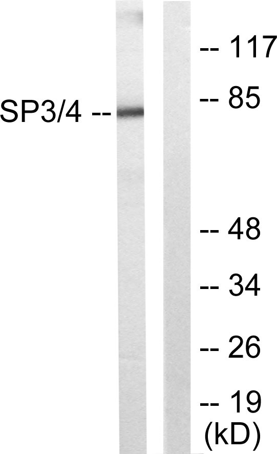
Cn|En

现货抗体产品库

SOCS-1 Rabbit Polyclonal Antibody

产品详情 相关文献 产品问答 相关产品

产品基本信息

产品货号
BD-PT4362
产品名称
SOCS-1 Rabbit Polyclonal Antibody
别名
SOCS1; SSI1; TIP3; Suppressor of cytokine signaling 1; SOCS-1; JAK-binding protein; JAB; STAT-induced STAT inhibitor 1; SSI-1; Tec-interacting protein 3; TIP-3
类别
常规抗体
基因名称
SOCS1
蛋白名称
Suppressor of cytokine signaling 1
推荐应用
WB
反应种属
Human,Mouse,Rat
浓度
1 mg/ml
存储缓冲液
Liquid in PBS containing 50% glycerol, 0.5% BSA and 0.02% New type preservative N.
Human Gene ID
8651
Human Gene Link
http://www.ncbi.nlm.nih.gov/sites/entrez?db=gene&term=8651
Human Swissprot No.
O15524
Human Swissprot Link
http://www.uniprot.org/uniprotkb/O15524/entry
Mouse Gene ID
12703
Mouse Gene Link
http://www.ncbi.nlm.nih.gov/sites/entrez?db=gene&term=12703
Mouse Swissprot No.
O35716
Mouse Swissprot Link
http://www.uniprot.org/uniprot/O35716
Rat Gene ID
252971
Rat Gene Link
http://www.ncbi.nlm.nih.gov/sites/entrez?db=gene&term=252971
Rat Swissprot No.
Q9QX78
Rat Swissprot Link
http://www.uniprot.org/uniprot/Q9QX78
免疫原
The antiserum was produced against synthesized peptide derived from human SOCS-1. AA range:49-98
特异性
SOCS-1 Polyclonal Antibody detects endogenous levels of SOCS-1 protein.
稀释度
IF 1:50-200 WB 1:500-2000, ELISA 1:10000-20000 IHC 1:50-300
预测分子量
38kD
运输及保存条件
-20°C/1 year
宿主
Polyclonal, Rabbit,IgG
背景介绍
This gene encodes a member of the STAT-induced STAT inhibitor (SSI), also known as suppressor of cytokine signaling (SOCS), family. SSI family members are cytokine-inducible negative regulators of cytokine signaling. The expression of this gene can be induced by a subset of cytokines, including IL2, IL3 erythropoietin (EPO), CSF2/GM-CSF, and interferon (IFN)-gamma. The protein encoded by this gene functions downstream of cytokine receptors, and takes part in a negative feedback loop to attenuate cytokine signaling. Knockout studies in mice suggested the role of this gene as a modulator of IFN-gamma action, which is required for normal postnatal growth and survival. [provided by RefSeq, Jul 2008],
组织表达
Expressed in all tissues with high expression in spleen, small intestine and peripheral blood leukocytes.
细胞定位
Nucleus . Cytoplasmic vesicle . Detected in perinuclear cytoplasmic vesicles upon interaction with FGFR3.
信号通路
Ubiquitin mediated proteolysis;Jak_STAT;Insulin_Receptor;Type II diabetes mellitus;
功能
domain:The ESS and SH2 domains are required for JAK phosphotyrosine binding. Further interaction with the KIR domain is necessary for signal and kinase inhibition.,domain:The SOCS box domain mediates the interaction with the Elongin BC complex, an adapter module in different E3 ubiquitin ligase complexes. The Elongin BC complex binding domain is also known as BC-box with the consensus [APST]-L-x(3)-C-x(3)-[AILV] and is part of the SOCS box.,function:SOCS family proteins form part of a classical negative feedback system that regulates cytokine signal transduction. SOCS1 is involved in negative regulation of cytokines that signal through the JAK/STAT3 pathway. Through binding to JAKs, inhibits their kinase activity. In vitro, also suppresses Tec protein-tyrosine activity. Appears to be a major regulator of signaling by interleukin 6 (IL6) and leukemia inhibitory factor (LIF). Regulates interferon-gamma mediated sensory neuron survival (By similarity). Probable substrate recognition component of an ECS (Elongin BC-CUL2/5-SOCS-box protein) E3 ubiquitin ligase complex which mediates the ubiquitination and subsequent proteasomal degradation of target proteins. Seems to recognize JAK2.,induction:By a subset of cytokines including those belonging to the interferon, interleukin and colony-stimulating factor families.,pathway:Protein modification; protein ubiquitination.,similarity:Contains 1 SH2 domain.,similarity:Contains 1 SOCS box domain.,subunit:Interacts with multiple activated signaling proteins of the tyrosine kinase signaling pathway including JAK family kinases, TEC, KIT, GRB2 and VAV. Binding to JAKs is mediated through the KIR and SH2 domains to a phosphorylated tyrosine residue within the JAK JH1 domain. Binds the SH3 domain of GRB2 via diproline determinants in the N-terminus, and the N-terminal regulatory domain of VAV (By similarity). Interacts with the Elongin BC complex (TCEB1 and TCEB2). Component of an ECS CBC(SOCS1) E3 ubiquitin-protein ligase complex which contains Elongin BC, CUL5, RBX1 and SOCS1 (By similarity). Interacts with TRIM8 (By similarity). Interacts with CUL2.,tissue specificity:Expressed in all tissues with high expression in spleen, small intestine and peripheral blood leukocytes.,
期货
现货
纯化
The antibody was affinity-purified from rabbit antiserum by affinity-chromatography using epitope-specific immunogen.

Western blot analysis of lysate from Jurkat cells, using SOCS-1 antibody.

Immunofluorescence analysis of mouse-liver tissue. 1,SOCS-1 Polyclonal Antibody(red) was diluted at 1:200(4°C,overnight). 2, Cy3 labled Secondary antibody was diluted at 1:300(room temperature, 50min).3, Picture B: DAPI(blue) 10min. Picture A:Target. Picture B: DAPI. Picture C: merge of A+B

Immunofluorescence analysis of mouse-liver tissue. 1,SOCS-1 Polyclonal Antibody(red) was diluted at 1:200(4°C,overnight). 2, Cy3 labled Secondary antibody was diluted at 1:300(room temperature, 50min).3, Picture B: DAPI(blue) 10min. Picture A:Target. Picture B: DAPI. Picture C: merge of A+B

Immunofluorescence analysis of mouse-lung tissue. 1,SOCS-1 Polyclonal Antibody(red) was diluted at 1:200(4°C,overnight). 2, Cy3 labled Secondary antibody was diluted at 1:300(room temperature, 50min).3, Picture B: DAPI(blue) 10min. Picture A:Target. Picture B: DAPI. Picture C: merge of A+B

Immunofluorescence analysis of mouse-lung tissue. 1,SOCS-1 Polyclonal Antibody(red) was diluted at 1:200(4°C,overnight). 2, Cy3 labled Secondary antibody was diluted at 1:300(room temperature, 50min).3, Picture B: DAPI(blue) 10min. Picture A:Target. Picture B: DAPI. Picture C: merge of A+B

Immunofluorescence analysis of mouse-kidney tissue. 1,SOCS-1 Polyclonal Antibody(red) was diluted at 1:200(4°C,overnight). 2, Cy3 labled Secondary antibody was diluted at 1:300(room temperature, 50min).3, Picture B: DAPI(blue) 10min. Picture A:Target. Picture B: DAPI. Picture C: merge of A+B

Immunofluorescence analysis of mouse-kidney tissue. 1,SOCS-1 Polyclonal Antibody(red) was diluted at 1:200(4°C,overnight). 2, Cy3 labled Secondary antibody was diluted at 1:300(room temperature, 50min).3, Picture B: DAPI(blue) 10min. Picture A:Target. Picture B: DAPI. Picture C: merge of A+B

Immunohistochemical analysis of paraffin-embedded Human-uterus tissue. 1,SOCS-1 Polyclonal Antibody was diluted at 1:200(4°C,overnight). 2, Sodium citrate pH 6.0 was used for antibody retrieval(>98°C,20min). 3,Secondary antibody was diluted at 1:200(room tempeRature, 30min). Negative control was used by secondary antibody only.

Immunohistochemical analysis of paraffin-embedded Human-uterus-cancer tissue. 1,SOCS-1 Polyclonal Antibody was diluted at 1:200(4°C,overnight). 2, Sodium citrate pH 6.0 was used for antibody retrieval(>98°C,20min). 3,Secondary antibody was diluted at 1:200(room tempeRature, 30min). Negative control was used by secondary antibody only.

Immunohistochemical analysis of paraffin-embedded Human-Tonsil tissue. 1,SOCS-1 Polyclonal Antibody was diluted at 1:200(4°C,overnight). 2, Sodium citrate pH 6.0 was used for antibody retrieval(>98°C,20min). 3,Secondary antibody was diluted at 1:200(room tempeRature, 30min). Negative control was used by secondary antibody only.

Immunohistochemical analysis of paraffin-embedded Human-liver tissue. 1,SOCS-1 Polyclonal Antibody was diluted at 1:200(4°C,overnight). 2, Sodium citrate pH 6.0 was used for antibody retrieval(>98°C,20min). 3,Secondary antibody was diluted at 1:200(room tempeRature, 30min). Negative control was used by secondary antibody only.

Immunohistochemical analysis of paraffin-embedded Human-liver-cancer tissue. 1,SOCS-1 Polyclonal Antibody was diluted at 1:200(4°C,overnight). 2, Sodium citrate pH 6.0 was used for antibody retrieval(>98°C,20min). 3,Secondary antibody was diluted at 1:200(room tempeRature, 30min). Negative control was used by secondary antibody only.

Immunohistochemical analysis of paraffin-embedded Human-lung tissue. 1,SOCS-1 Polyclonal Antibody was diluted at 1:200(4°C,overnight). 2, Sodium citrate pH 6.0 was used for antibody retrieval(>98°C,20min). 3,Secondary antibody was diluted at 1:200(room tempeRature, 30min). Negative control was used by secondary antibody only.

Immunohistochemical analysis of paraffin-embedded Human-stomach tissue. 1,SOCS-1 Polyclonal Antibody was diluted at 1:200(4°C,overnight). 2, Sodium citrate pH 6.0 was used for antibody retrieval(>98°C,20min). 3,Secondary antibody was diluted at 1:200(room tempeRature, 30min). Negative control was used by secondary antibody only.

Immunohistochemical analysis of paraffin-embedded Human-stomach-cancer tissue. 1,SOCS-1 Polyclonal Antibody was diluted at 1:200(4°C,overnight). 2, Sodium citrate pH 6.0 was used for antibody retrieval(>98°C,20min). 3,Secondary antibody was diluted at 1:200(room tempeRature, 30min). Negative control was used by secondary antibody only.

Immunohistochemical analysis of paraffin-embedded Rat-heart tissue. 1,SOCS-1 Polyclonal Antibody was diluted at 1:200(4°C,overnight). 2, Sodium citrate pH 6.0 was used for antibody retrieval(>98°C,20min). 3,Secondary antibody was diluted at 1:200(room tempeRature, 30min). Negative control was used by secondary antibody only.

Immunohistochemical analysis of paraffin-embedded Rat-testis tissue. 1,SOCS-1 Polyclonal Antibody was diluted at 1:200(4°C,overnight). 2, Sodium citrate pH 6.0 was used for antibody retrieval(>98°C,20min). 3,Secondary antibody was diluted at 1:200(room tempeRature, 30min). Negative control was used by secondary antibody only.

Immunohistochemical analysis of paraffin-embedded Rat-liver tissue. 1,SOCS-1 Polyclonal Antibody was diluted at 1:200(4°C,overnight). 2, Sodium citrate pH 6.0 was used for antibody retrieval(>98°C,20min). 3,Secondary antibody was diluted at 1:200(room tempeRature, 30min). Negative control was used by secondary antibody only.

Immunohistochemical analysis of paraffin-embedded Rat-kidney tissue. 1,SOCS-1 Polyclonal Antibody was diluted at 1:200(4°C,overnight). 2, Sodium citrate pH 6.0 was used for antibody retrieval(>98°C,20min). 3,Secondary antibody was diluted at 1:200(room tempeRature, 30min). Negative control was used by secondary antibody only.

Immunohistochemical analysis of paraffin-embedded Rat-brain tissue. 1,SOCS-1 Polyclonal Antibody was diluted at 1:200(4°C,overnight). 2, Sodium citrate pH 6.0 was used for antibody retrieval(>98°C,20min). 3,Secondary antibody was diluted at 1:200(room tempeRature, 30min). Negative control was used by secondary antibody only.

Immunohistochemical analysis of paraffin-embedded Mouse-heart tissue. 1,SOCS-1 Polyclonal Antibody was diluted at 1:200(4°C,overnight). 2, Sodium citrate pH 6.0 was used for antibody retrieval(>98°C,20min). 3,Secondary antibody was diluted at 1:200(room tempeRature, 30min). Negative control was used by secondary antibody only.

Immunohistochemical analysis of paraffin-embedded Mouse-testis tissue. 1,SOCS-1 Polyclonal Antibody was diluted at 1:200(4°C,overnight). 2, Sodium citrate pH 6.0 was used for antibody retrieval(>98°C,20min). 3,Secondary antibody was diluted at 1:200(room tempeRature, 30min). Negative control was used by secondary antibody only.

Immunohistochemical analysis of paraffin-embedded Mouse-liver tissue. 1,SOCS-1 Polyclonal Antibody was diluted at 1:200(4°C,overnight). 2, Sodium citrate pH 6.0 was used for antibody retrieval(>98°C,20min). 3,Secondary antibody was diluted at 1:200(room tempeRature, 30min). Negative control was used by secondary antibody only.

Immunohistochemical analysis of paraffin-embedded Mouse-lung tissue. 1,SOCS-1 Polyclonal Antibody was diluted at 1:200(4°C,overnight). 2, Sodium citrate pH 6.0 was used for antibody retrieval(>98°C,20min). 3,Secondary antibody was diluted at 1:200(room tempeRature, 30min). Negative control was used by secondary antibody only.

Immunohistochemical analysis of paraffin-embedded Mouse-kidney tissue. 1,SOCS-1 Polyclonal Antibody was diluted at 1:200(4°C,overnight). 2, Sodium citrate pH 6.0 was used for antibody retrieval(>98°C,20min). 3,Secondary antibody was diluted at 1:200(room tempeRature, 30min). Negative control was used by secondary antibody only.

Western Blot analysis of various cells using SOCS-1 Polyclonal Antibody diluted at 1:2000

相关文献

产品问答

相关产品

免责声明| 法律支持| 联系方式

市场:027-65023363   行政/人事:027-62439686   邮箱:marketing@brainvta.com  

销售总监:张经理  18995532642  华东区:陈经理 18013970337   华南区:王经理 13100653525   华中/西区:杨经理 18186518905   华北区:张经理 18893721749

地址:中国武汉东湖高新区光谷七路128号中科开物产业园1号楼

Copyright © 武汉枢密脑科学技术有限公司. All RIGHTS RESERVED.
鄂ICP备2021009124号 DIGITAL BY VTHINK