产品名称
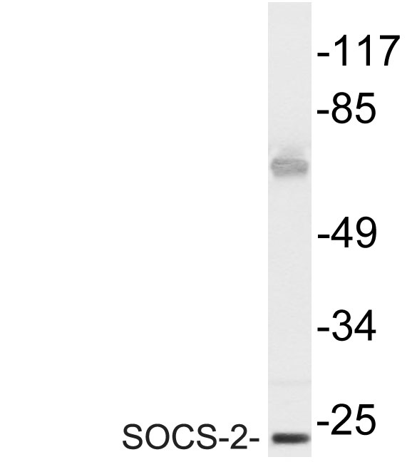
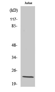
SOCS-2 Rabbit Polyclonal Antibody
别名
SOCS2; CIS2; SSI2; STATI2; Suppressor of cytokine signaling 2; SOCS-2; Cytokine-inducible SH2 protein 2; CIS-2; STAT-induced STAT inhibitor 2; SSI-2
蛋白名称
Suppressor of cytokine signaling 2
存储缓冲液
Liquid in PBS containing 50% glycerol, 0.5% BSA and 0.02% New type preservative N.
Human Gene Link
http://www.ncbi.nlm.nih.gov/sites/entrez?db=gene&term=8835
Human Swissprot No.
O14508
Human Swissprot Link
http://www.uniprot.org/uniprotkb/O14508/entry
Mouse Gene Link
http://www.ncbi.nlm.nih.gov/sites/entrez?db=gene&term=216233
Mouse Swissprot No.
O35717
Mouse Swissprot Link
http://www.uniprot.org/uniprot/O35717
Rat Gene Link
http://www.ncbi.nlm.nih.gov/sites/entrez?db=gene&term=84607
Rat Swissprot Link
http://www.uniprot.org/uniprot/O88582
免疫原
The antiserum was produced against synthesized peptide derived from human SOCS-2. AA range:18-67
特异性
SOCS-2 Polyclonal Antibody detects endogenous levels of SOCS-2 protein.
宿主
Polyclonal, Rabbit,IgG
背景介绍
This gene encodes a member of the suppressor of cytokine signaling (SOCS) family. SOCS family members are cytokine-inducible negative regulators of cytokine receptor signaling via the Janus kinase/signal transducer and activation of transcription pathway (the JAK/STAT pathway). SOCS family proteins interact with major molecules of signaling complexes to block further signal transduction, in part, by proteasomal depletion of receptors or signal-transducing proteins via ubiquitination. The expression of this gene can be induced by a subset of cytokines, including erythropoietin, GM-CSF, IL10, interferon (IFN)-gamma and by cytokine receptors such as growth horomone receptor. The protein encoded by this gene interacts with the cytoplasmic domain of insulin-like growth factor-1 receptor (IGF1R) and is thought to be involved in the regulation of IGF1R mediated cell signaling. This gene has
组织表达
High expression in heart, placenta, lung, kidney and prostate. Predominantly expressed in pulmonary epithelia cells, specifically type II pneumocytes.
信号通路
Jak_STAT;Insulin_Receptor;Type II diabetes mellitus;
功能
domain:The SOCS box domain mediates the interaction with the Elongin BC complex, an adapter module in different E3 ubiquitin ligase complexes.,function:SOCS family proteins form part of a classical negative feedback system that regulates cytokine signal transduction. SOCS2 appears to be a negative regulator in the growth hormone/IGF1 signaling pathway. Probable substrate recognition component of a SCF-like ECS (Elongin BC-CUL2/5-SOCS-box protein) E3 ubiquitin-protein ligase complex which mediates the ubiquitination and subsequent proteasomal degradation of target proteins.,induction:By a subset of cytokines, including erythropoietin and granulocyte-macrophage colony stimulating factor (GM-CSF).,pathway:Protein modification; protein ubiquitination.,similarity:Contains 1 SH2 domain.,similarity:Contains 1 SOCS box domain.,subunit:Interacts with IGF1 receptor, prolactin receptor and growth hormone (GH) receptor. Associates with the Elongin BC complex.,tissue specificity:High expression in heart, placenta, lung, kidney and prostate.,
纯化
The antibody was affinity-purified from rabbit antiserum by affinity-chromatography using epitope-specific immunogen.