产品名称
SMVT Rabbit Polyclonal Antibody
别名
SLC5A6; SMVT; Sodium-dependent multivitamin transporter; Na(+)-dependent multivitamin transporter; Solute carrier family 5 member 6
蛋白名称
Sodium-dependent multivitamin transporter
存储缓冲液
Liquid in PBS containing 50% glycerol, 0.5% BSA and 0.02% New type preservative N.
Human Gene Link
http://www.ncbi.nlm.nih.gov/sites/entrez?db=gene&term=8884
Human Swissprot No.
Q9Y289
Human Swissprot Link
http://www.uniprot.org/uniprotkb/Q9Y289/entry
Mouse Swissprot No.
Q5U4D8
Mouse Swissprot Link
http://www.uniprot.org/uniprot/Q5U4D8
免疫原
The antiserum was produced against synthesized peptide derived from human SLC5A6. AA range:551-600
特异性
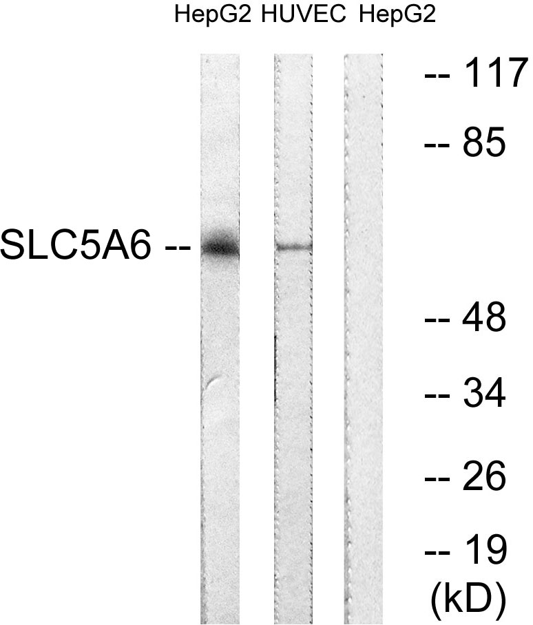
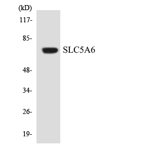
SMVT Polyclonal Antibody detects endogenous levels of SMVT protein.
宿主
Polyclonal, Rabbit,IgG
背景介绍
function:Transports pantothenate, biotin and lipoate in the presence of sodium.,similarity:Belongs to the sodium:solute symporter (SSF) (TC 2.A.21) family.,
组织表达
Expressed in microvessels of the brain (at protein level) (PubMed:25809983). Expressed in heart, brain, placenta, lung, liver, skeletal muscle, kidney, and pancreas (PubMed:10329687).
细胞定位
Cell membrane ; Multi-pass membrane protein .; Cell membrane ; Multi-pass membrane protein . (Microbial infection) Exposure to E.coli lipopolysaccharides leeds to reduced cell membrane localization. .
功能
function:Transports pantothenate, biotin and lipoate in the presence of sodium.,similarity:Belongs to the sodium:solute symporter (SSF) (TC 2.A.21) family.,
纯化
The antibody was affinity-purified from rabbit antiserum by affinity-chromatography using epitope-specific immunogen.