产品名称
PRDM3 Rabbit Polyclonal Antibody
别名
MECOM; MDS1; MDS1 and EVI1 complex locus protein MDS1; Myelodysplasia syndrome 1 protein; Myelodysplasia syndrome-associated protein 1
蛋白名称
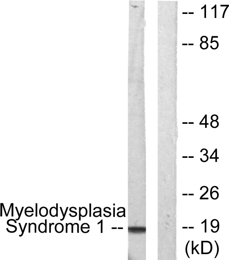
MDS1 and EVI1 complex locus protein MDS1
存储缓冲液
Liquid in PBS containing 50% glycerol, 0.5% BSA and 0.02% New type preservative N.
Human Gene Link
http://www.ncbi.nlm.nih.gov/sites/entrez?db=gene&term=4197
Human Swissprot No.
Q13465
Human Swissprot Link
http://www.uniprot.org/uniprotkb/Q13465/entry
Mouse Swissprot No.
Q9Z1L8
Mouse Swissprot Link
http://www.uniprot.org/uniprot/Q9Z1L8
免疫原
The antiserum was produced against synthesized peptide derived from human MECOM. AA range:1-50
特异性
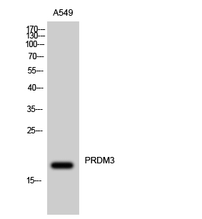
PRDM3 Polyclonal Antibody detects endogenous levels of PRDM3 protein.
宿主
Polyclonal, Rabbit,IgG
背景介绍
The protein encoded by this gene is a transcriptional regulator and oncoprotein that may be involved in hematopoiesis, apoptosis, development, and cell differentiation and proliferation. The encoded protein can interact with CTBP1, SMAD3, CREBBP, KAT2B, MAPK8, and MAPK9. This gene can undergo translocation with the AML1 gene, resulting in overexpression of this gene and the onset of leukemia. Several transcript variants encoding a few different isoforms have been found for this gene. [provided by RefSeq, Mar 2011],
细胞定位
histone deacetylase complex,nucleus,nucleoplasm,cytoplasm,Golgi apparatus,cytosol,aggresome,nuclear speck,intracellular membrane-bounded organelle,
信号通路
MAPK_ERK_Growth;MAPK_G_Protein;Pathways in cancer;Chronic myeloid leukemia;
功能
disease:A chromosomal aberration involving EVI1 is a cause of chronic myelogenous leukemia (CML). Translocation t(3;21)(q26;q22) with RUNX1/AML1.,disease:A chromosomal aberration involving MDS1 is found in a form of acute myeloid leukemia (AML). Translocation t(3;21) with AML1.,miscellaneous:Can be produced either as a separate transcript and as a normal fusion transcript with EVI1.,PTM:Phosphorylated upon DNA damage, probably by ATM or ATR.,similarity:Contains 10 C2H2-type zinc fingers.,subunit:May interact with CTBP1.,
纯化
The antibody was affinity-purified from rabbit antiserum by affinity-chromatography using epitope-specific immunogen.