产品名称
PFKM Rabbit Polyclonal Antibody
别名
PFKM; PFKX; 6-phosphofructokinase; muscle type; Phosphofructo-1-kinase isozyme A; PFK-A; Phosphofructokinase-M; Phosphofructokinase 1; Phosphohexokinase
蛋白名称
6-phosphofructokinase muscle type
存储缓冲液
Liquid in PBS containing 50% glycerol, 0.5% BSA and 0.02% New type preservative N.
Human Gene Link
http://www.ncbi.nlm.nih.gov/sites/entrez?db=gene&term=5213
Human Swissprot No.
P08237
Human Swissprot Link
http://www.uniprot.org/uniprotkb/P08237/entry
Mouse Gene Link
http://www.ncbi.nlm.nih.gov/sites/entrez?db=gene&term=18642
Mouse Swissprot No.
P47857
Mouse Swissprot Link
http://www.uniprot.org/uniprot/P47857
Rat Gene Link
http://www.ncbi.nlm.nih.gov/sites/entrez?db=gene&term=65152
Rat Swissprot Link
http://www.uniprot.org/uniprot/P47858
免疫原
The antiserum was produced against synthesized peptide derived from human PFK-1. AA range:320-369
特异性
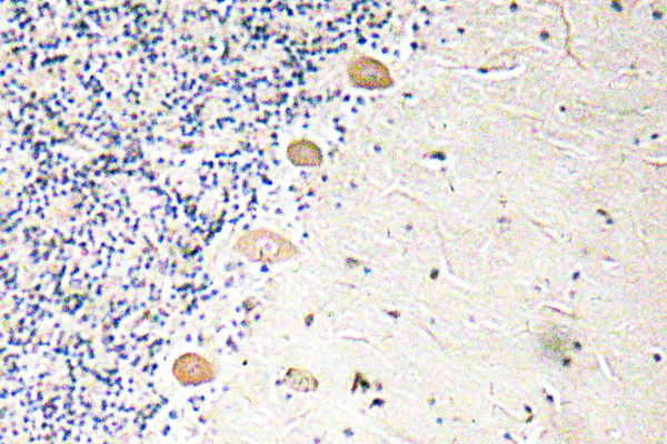
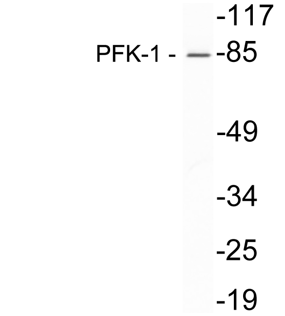
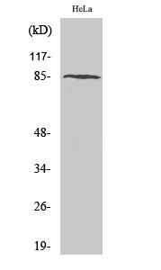
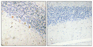
PFKM Polyclonal Antibody detects endogenous levels of PFKM protein.
宿主
Polyclonal, Rabbit,IgG
背景介绍
Three phosphofructokinase isozymes exist in humans: muscle, liver and platelet. These isozymes function as subunits of the mammalian tetramer phosphofructokinase, which catalyzes the phosphorylation of fructose-6-phosphate to fructose-1,6-bisphosphate. Tetramer composition varies depending on tissue type. This gene encodes the muscle-type isozyme. Mutations in this gene have been associated with glycogen storage disease type VII, also known as Tarui disease. Alternatively spliced transcript variants have been described.[provided by RefSeq, Nov 2009],
组织表达
Brain,Liver,Muscle,Skeletal muscle,Thymus,
信号通路
Glycolysis / Gluconeogenesis;Pentose phosphate pathway;Fructose and mannose metabolism;Galactose metabolism;
功能
catalytic activity:ATP + D-fructose 6-phosphate = ADP + D-fructose 1,6-bisphosphate.,cofactor:Magnesium.,disease:Defects in PFKM are the cause of glycogen storage disease type 7 (GSD7) [MIM:232800]; also known as Tarui disease. GSD7 is an autosomal recessive disorder characterized by exercise intolerance with associated nausea and vomiting. Short bursts of intense activity are particularly difficult. Severe muscle cramps and myoglobinuria develop after vigorous exercise. Most patients obtain a "second wind" when the onset of exercise is followed by a brief rest period. In time patients adjust their activity level and are well compensated.,enzyme regulation:Allosteric enzyme activated by ADP, AMP, or fructose bisphosphate and inhibited by ATP or citrate.,miscellaneous:In human PFK exists as a system of 3 types of subunits, PFKM (muscle), PFKL (liver) and PFKP (platelet) isoenzymes.,pathway:Carbohydrate degradation; glycolysis; D-glyceraldehyde 3-phosphate and glycerone phosphate from D-glucose: step 3/4.,similarity:Belongs to the phosphofructokinase family. Two domains subfamily.,subunit:Tetramer. Muscle is M4, liver is L4, and red cell is M3L, M2L2, or ML3.,
纯化
The antibody was affinity-purified from rabbit antiserum by affinity-chromatography using epitope-specific immunogen.