产品名称
Pepsin A Rabbit Polyclonal Antibody
别名
Pepsin A; PGA3; PGA4; PGA5
存储缓冲液
Liquid in PBS containing 50% glycerol, 0.5% BSA and 0.02% New type preservative N.
Human Gene ID
5222/643847
Human Gene Link
http://www.ncbi.nlm.nih.gov/sites/entrez?db=gene&term=5222
Human Swissprot No.
P00790
Human Swissprot Link
http://www.uniprot.org/uniprotkb/P00790/entry
免疫原
The antiserum was produced against synthesized peptide derived from human Pepsin A. AA range:258-307
特异性
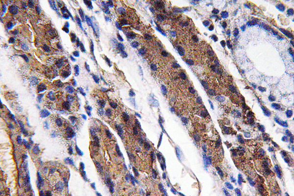
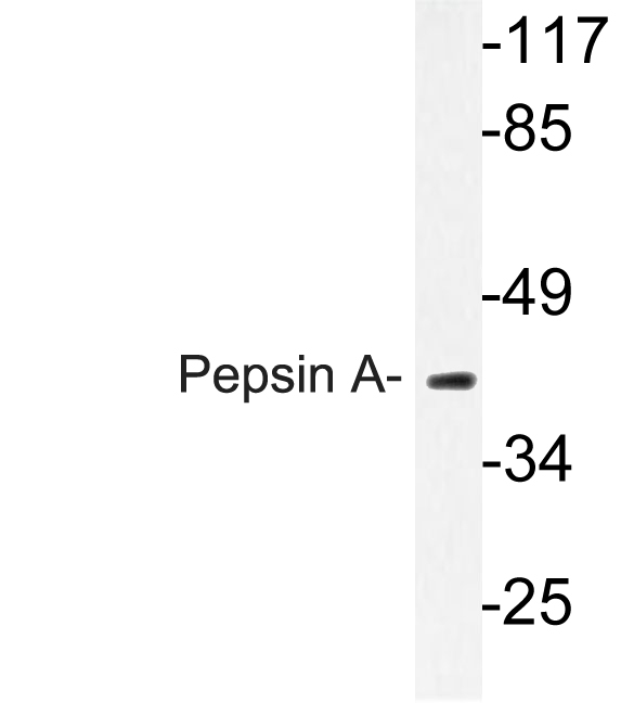
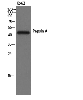
Pepsin A Polyclonal Antibody detects endogenous levels of Pepsin A protein.
宿主
Polyclonal, Rabbit,IgG
背景介绍
This gene encodes a protein precursor of the digestive enzyme pepsin, a member of the peptidase A1 family of endopeptidases. The encoded precursor is secreted by gastric chief cells and undergoes autocatalytic cleavage in acidic conditions to form the active enzyme, which functions in the digestion of dietary proteins. This gene is found in a cluster of related genes on chromosome 11, each of which encodes one of multiple pepsinogens. Pepsinogen levels in serum may serve as a biomarker for atrophic gastritis and gastric cancer. [provided by RefSeq, Jul 2015],
组织表达
Brain,Colon,Kidney,Placenta,Skeletal muscle,
细胞定位
extracellular exosome,multivesicular body lumen,
功能
catalytic activity:Preferential cleavage: hydrophobic, preferably aromatic, residues in P1 and P1' positions. Cleaves 1-Phe-|-Val-2, 4-Gln-|-His-5, 13-Glu-|-Ala-14, 14-Ala-|-Leu-15, 15-Leu-|-Tyr-16, 16-Tyr-|-Leu-17, 23-Gly-|-Phe-24, 24-Phe-|-Phe-25 and 25-Phe-|-Tyr-26 bonds in the B chain of insulin.,function:Shows particularly broad specificity; although bonds involving phenylalanine and leucine are preferred, many others are also cleaved to some extent.,similarity:Belongs to the peptidase A1 family.,
纯化
The antibody was affinity-purified from rabbit antiserum by affinity-chromatography using epitope-specific immunogen.