产品名称
NRBF-2 Rabbit Polyclonal Antibody
别名
NRBF2; COPR; Nuclear receptor-binding factor 2; NRBF-2; Comodulator of PPAR and RXR
蛋白名称
Nuclear receptor-binding factor 2
存储缓冲液
Liquid in PBS containing 50% glycerol, 0.5% BSA and 0.02% New type preservative N.
Human Gene Link
http://www.ncbi.nlm.nih.gov/sites/entrez?db=gene&term=29982
Human Swissprot No.
Q96F24
Human Swissprot Link
http://www.uniprot.org/uniprotkb/Q96F24/entry
Mouse Gene Link
http://www.ncbi.nlm.nih.gov/sites/entrez?db=gene&term=641340
Mouse Swissprot No.
Q8VCQ3
Mouse Swissprot Link
http://www.uniprot.org/uniprot/Q8VCQ3
Rat Gene Link
http://www.ncbi.nlm.nih.gov/sites/entrez?db=gene&term=58839
Rat Swissprot Link
http://www.uniprot.org/uniprot/Q9QYK3
免疫原
The antiserum was produced against synthesized peptide derived from human NRBF2. AA range:140-189
特异性
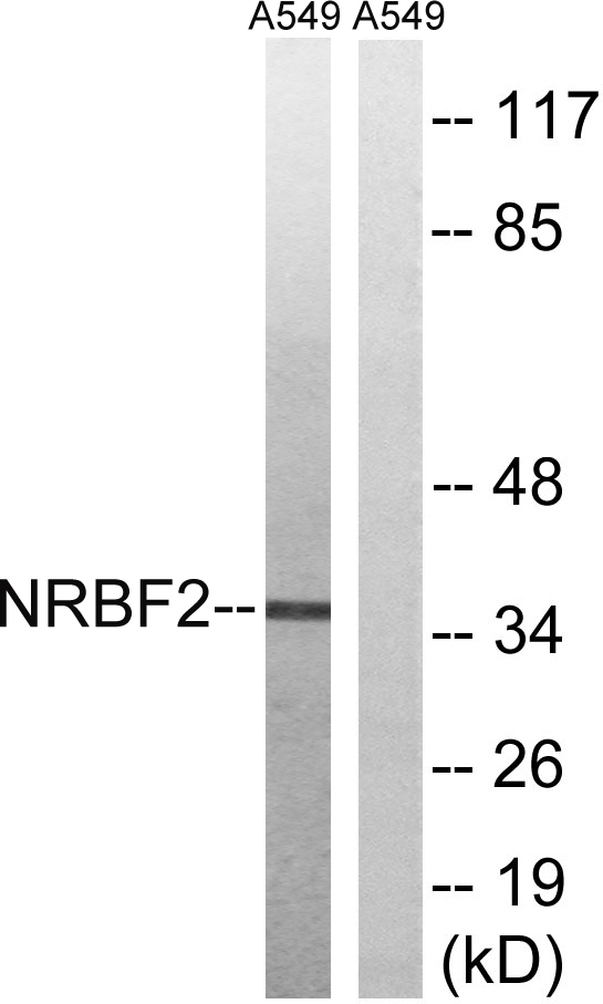
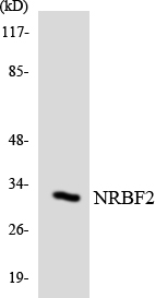
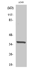
NRBF-2 Polyclonal Antibody detects endogenous levels of NRBF-2 protein.
宿主
Polyclonal, Rabbit,IgG
背景介绍
function:May modulate transcriptional activation by target nuclear receptors. Can act as transcriptional activator (in vitro).,subunit:Interacts with PPARA, PPARD and PPARG. Interacts with RARA, RARG and RXRA in the presence of bound ligand.,tissue specificity:Detected in keratinocytes, liver and placenta.,
组织表达
Detected in keratinocytes, liver and placenta (PubMed:15610520). Expressed in a subset of cells in pediatric medulloblastoma (PubMed:18619852).
细胞定位
Nucleus . Cytoplasm . Cytoplasmic vesicle . Cytoplasmic vesicle, autophagosome .
功能
function:May modulate transcriptional activation by target nuclear receptors. Can act as transcriptional activator (in vitro).,subunit:Interacts with PPARA, PPARD and PPARG. Interacts with RARA, RARG and RXRA in the presence of bound ligand.,tissue specificity:Detected in keratinocytes, liver and placenta.,
纯化
The antibody was affinity-purified from rabbit antiserum by affinity-chromatography using epitope-specific immunogen.