产品名称
KCNG2 Rabbit Polyclonal Antibody
别名
KCNG2; KCNF2; Potassium voltage-gated channel subfamily G member 2; Cardiac potassium channel subunit; Voltage-gated potassium channel subunit Kv6.2
蛋白名称
Potassium voltage-gated channel subfamily G member 2
存储缓冲液
Liquid in PBS containing 50% glycerol, 0.5% BSA and 0.02% New type preservative N.
Human Gene Link
http://www.ncbi.nlm.nih.gov/sites/entrez?db=gene&term=26251
Human Swissprot No.
Q9UJ96
Human Swissprot Link
http://www.uniprot.org/uniprotkb/Q9UJ96/entry
Rat Gene Link
http://www.ncbi.nlm.nih.gov/sites/entrez?db=gene&term=307234
Rat Swissprot Link
http://www.uniprot.org/uniprot/Q9QYU3
免疫原
The antiserum was produced against synthesized peptide derived from human KCNG2. AA range:321-370
特异性
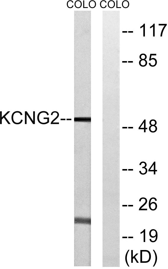
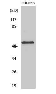
KCNG2 Polyclonal Antibody detects endogenous levels of KCNG2 protein.
宿主
Polyclonal, Rabbit,IgG
背景介绍
Voltage-gated potassium (Kv) channels represent the most complex class of voltage-gated ion channels from both functional and structural standpoints. Their diverse functions include regulating neurotransmitter release, heart rate, insulin secretion, neuronal excitability, epithelial electrolyte transport, smooth muscle contraction, and cell volume. This gene encodes a member of the potassium channel, voltage-gated, subfamily G. This member is a gamma subunit of the voltage-gated potassium channel. The delayed-rectifier type channels containing this subunit may contribute to cardiac action potential repolarization. [provided by RefSeq, Jul 2008],
组织表达
Highly expressed in heart, liver, skeletal muscle, kidney and pancreas. Detected at low levels in brain, lung and placenta.
细胞定位
Membrane; Multi-pass membrane protein.
功能
domain:The segment S4 is probably the voltage-sensor and is characterized by a series of positively charged amino acids at every third position.,function:Potassium channel subunit. Modulates channel activity by shifting the threshold and the half-maximal activation to more negative values.,miscellaneous:Heterodimers with KCNB1 are highly sensitive to inhibition by tetraethylammonium (TEA) and propafenone.,similarity:Belongs to the potassium channel family. G subfamily.,subunit:Heterodimer with KCNB1. Does not form homomultimers.,tissue specificity:Highly expressed in heart, liver, skeletal muscle, kidney and pancreas. Detected at low levels in brain, lung and placenta.,
纯化
The antibody was affinity-purified from rabbit antiserum by affinity-chromatography using epitope-specific immunogen.