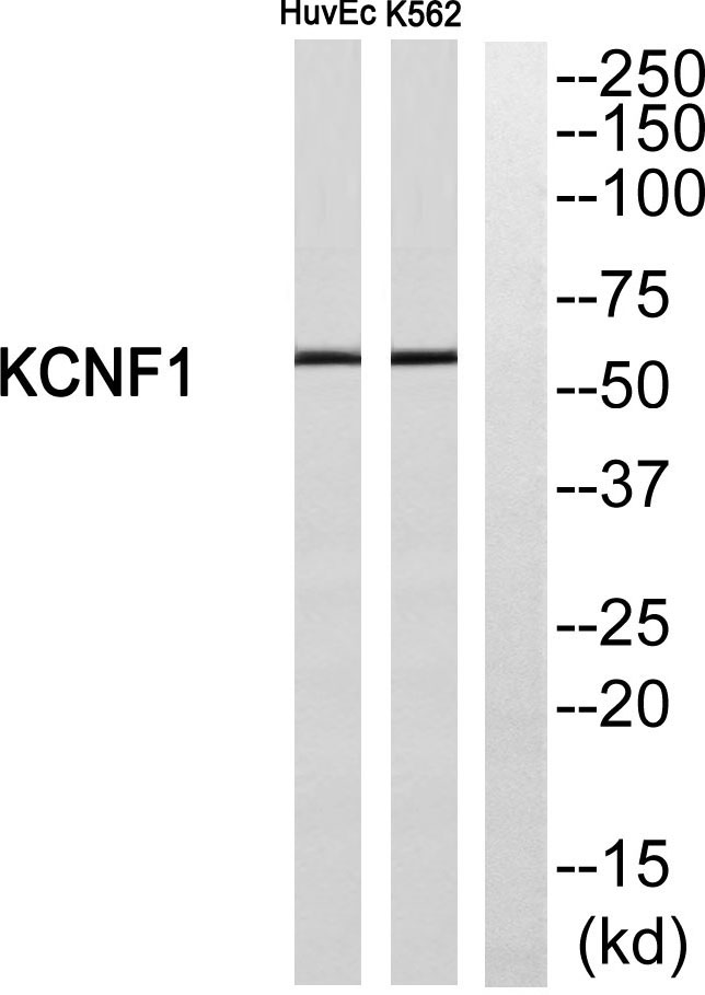
Cn|En

现货抗体产品库

JIP-2 Rabbit Polyclonal Antibody

产品详情 相关文献 产品问答 相关产品

产品基本信息

产品货号
BD-PT2435
产品名称
JIP-2 Rabbit Polyclonal Antibody
别名
MAPK8IP2; IB2; JIP2; PRKM8IPL; C-Jun-amino-terminal kinase-interacting protein 2; JIP-2; JNK-interacting protein 2; Islet-brain-2; IB-2; JNK MAP kinase scaffold protein 2; Mitogen-activated protein kinase 8-interacting protein 2
类别
常规抗体
基因名称
MAPK8IP2
蛋白名称
C-Jun-amino-terminal kinase-interacting protein 2
推荐应用
WB
反应种属
Human,Mouse,Rat
浓度
1 mg/ml
存储缓冲液
Liquid in PBS containing 50% glycerol, 0.5% BSA and 0.02% New type preservative N.
Human Gene ID
23542
Human Gene Link
http://www.ncbi.nlm.nih.gov/sites/entrez?db=gene&term=23542
Human Swissprot No.
Q13387
Human Swissprot Link
http://www.uniprot.org/uniprotkb/Q13387/entry
Mouse Gene ID
60597
Mouse Gene Link
http://www.ncbi.nlm.nih.gov/sites/entrez?db=gene&term=60597
Mouse Swissprot No.
Q9ERE9
Mouse Swissprot Link
http://www.uniprot.org/uniprot/Q9ERE9
免疫原
The antiserum was produced against synthesized peptide derived from human JIP2. AA range:581-630
特异性
JIP-2 Polyclonal Antibody detects endogenous levels of JIP-2 protein.
稀释度
WB 1:500 - 1:2000. IHC 1:100 - 1:300. IF 1:200 - 1:1000. ELISA: 1:20000. Not yet tested in other applications.
预测分子量
87kD
运输及保存条件
-20°C/1 year
宿主
Polyclonal, Rabbit,IgG
背景介绍
The protein encoded by this gene is closely related to MAPK8IP1/IB1/JIP-1, a scaffold protein that is involved in the c-Jun amino-terminal kinase signaling pathway. This protein is expressed in brain and pancreatic cells. It has been shown to interact with, and regulate the activity of MAPK8/JNK1, and MAP2K7/MKK7 kinases. This protein thus is thought to function as a regulator of signal transduction by protein kinase cascade in brain and pancreatic beta-cells. [provided by RefSeq, Feb 2014],
组织表达
Expressed mainly in the brain and pancreas, including insulin-secreting cells. In the nervous system, more abundantly expressed in the cerebellum, pituitary gland, occipital lobe and the amygdala. Also expressed in fetal brain. Very low levels found in uterus, ovary, prostate, colon, testis, adrenal gland, thyroid gland and salivary gland.
细胞定位
Cytoplasm . Accumulates in cell surface projections. .
信号通路
MAPK_ERK_Growth;MAPK_G_Protein;
功能
alternative products:Experimental confirmation may be lacking for some isoforms,function:The JNK-interacting protein (JIP) group of scaffold proteins selectively mediates JNK signaling by aggregating specific components of the MAPK cascade to form a functional JNK signaling module. JIP2 inhibits IL1 beta-induced apoptosis in insulin-secreting cells. May function as a regulator of vesicle transport, through interations with the JNK-signaling components and motor proteins.,similarity:Belongs to the JIP scaffold family.,similarity:Contains 1 PID domain.,similarity:Contains 1 SH3 domain.,subcellular location:Accumulates in cell surface projections.,subunit:Forms homo- or heterooligomeric complexes. Binds specific components of the JNK signaling pathway namely JNK, MAPKK7 and MLK2, MLK3 and DLK. Also binds the proline-rich domain-containing splice variant of apolipoprotein E receptor 2 (ApoER2). Binds the cytoplasmic tails of LRP1 and LRP2 (Megalin). Binds the TPR motif-containing C-terminal of kinesin light chain, Klc1, pre-assembled MAPK8IP1 scaffolding complexes are then transported as a cargo of kinesin, to the required subcellular location (By similarity). Interacts with the cytoplasmic domain of APP.,tissue specificity:Expressed mainly in the brain and pancreas, including insulin-secreting cells. In the nervous system, more abundantly expressed in the cerebellum, pituitary gland, occipital lobe and the amygdala. Also expressed in fetal brain. Very low levels found in uterus, ovary, prostate, colon, testis, adrenal gland, thyroid gland and salivary gland.,
期货
现货
纯化
The antibody was affinity-purified from rabbit antiserum by affinity-chromatography using epitope-specific immunogen.

Immunofluorescence analysis of HeLa cells, using JIP2 Antibody. The picture on the right is blocked with the synthesized peptide.

Immunohistochemistry analysis of paraffin-embedded human brain tissue, using JIP2 Antibody. The picture on the right is blocked with the synthesized peptide.

Western blot analysis of lysates from mouse brain, using JIP2 Antibody. The lane on the right is blocked with the synthesized peptide.

相关文献

产品问答

相关产品

免责声明| 法律支持| 联系方式

市场:027-65023363   行政/人事:027-62439686   邮箱:marketing@brainvta.com  

销售总监:张经理  18995532642  华东区:陈经理 18013970337   华南区:王经理 13100653525   华中/西区:杨经理 18186518905   华北区:张经理 18893721749

地址:中国武汉东湖高新区光谷七路128号中科开物产业园1号楼

Copyright © 武汉枢密脑科学技术有限公司. All RIGHTS RESERVED.
鄂ICP备2021009124号 DIGITAL BY VTHINK