产品名称
JTB Rabbit Polyclonal Antibody
别名
JTB; HSPC222; Protein JTB; Jumping translocation breakpoint protein; Prostate androgen-regulated protein; PAR protein
存储缓冲液
Liquid in PBS containing 50% glycerol, 0.5% BSA and 0.02% New type preservative N.
Human Gene Link
http://www.ncbi.nlm.nih.gov/sites/entrez?db=gene&term=10899
Human Swissprot No.
O76095
Human Swissprot Link
http://www.uniprot.org/uniprotkb/O76095/entry
Mouse Swissprot No.
O88824
Mouse Swissprot Link
http://www.uniprot.org/uniprot/O88824
免疫原
The antiserum was produced against synthesized peptide derived from human JTB. AA range:10-59
特异性
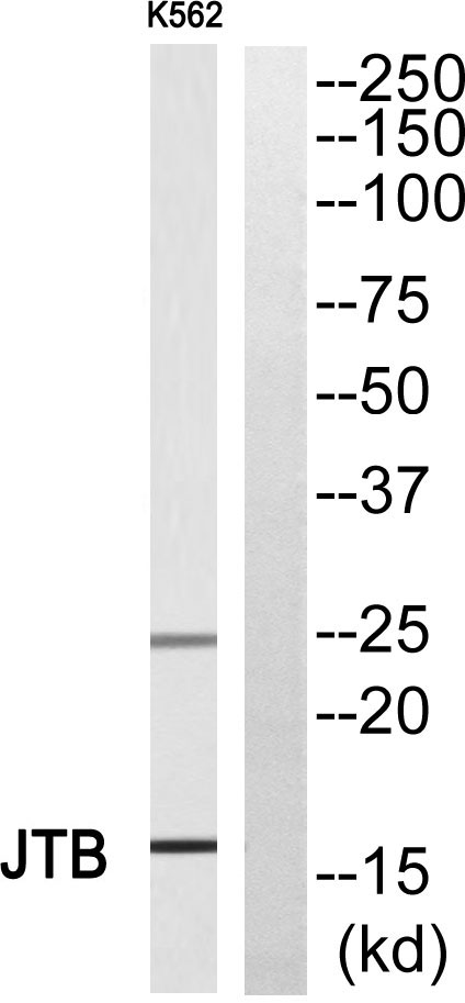
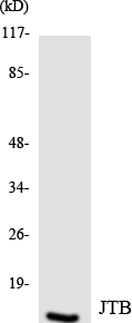
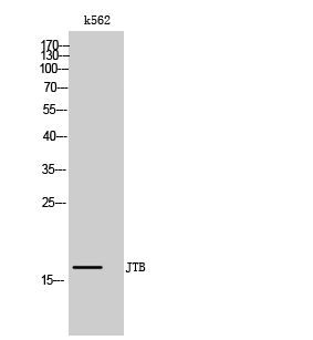
JTB Polyclonal Antibody detects endogenous levels of JTB protein.
宿主
Polyclonal, Rabbit,IgG
背景介绍
similarity:Belongs to the JTB family.,tissue specificity:Expressed in all normal human tissues studied but overexpressed in most of their malignant counterparts.,
组织表达
Ubiquitous. Expressed in all normal human tissues studied but overexpressed or underexpressed in many of their malignant counterparts.
细胞定位
Membrane ; Single-pass type I membrane protein . Mitochondrion . Cytoplasm. Cytoplasm, cytoskeleton, microtubule organizing center, centrosome. Cytoplasm, cytoskeleton, spindle. Detected at the centrosome and along spindle fibers during prophase and metaphase. Detected at the midbody during telophase.
功能
similarity:Belongs to the JTB family.,tissue specificity:Expressed in all normal human tissues studied but overexpressed in most of their malignant counterparts.,
纯化
The antibody was affinity-purified from rabbit antiserum by affinity-chromatography using epitope-specific immunogen.