产品名称
JIP-1 Rabbit Polyclonal Antibody
别名
MAPK8IP1; IB1; JIP1; PRKM8IP; C-Jun-amino-terminal kinase-interacting protein 1; JIP-1; JNK-interacting protein 1; Islet-brain 1; IB-1; JNK MAP kinase scaffold protein 1; Mitogen-activated protein kinase 8-interacting protein 1
蛋白名称
C-Jun-amino-terminal kinase-interacting protein 1
存储缓冲液
Liquid in PBS containing 50% glycerol, 0.5% BSA and 0.02% New type preservative N.
Human Gene Link
http://www.ncbi.nlm.nih.gov/sites/entrez?db=gene&term=9479
Human Swissprot No.
Q9UQF2
Human Swissprot Link
http://www.uniprot.org/uniprotkb/Q9UQF2/entry
Mouse Gene Link
http://www.ncbi.nlm.nih.gov/sites/entrez?db=gene&term=19099
Mouse Swissprot No.
Q9WVI9
Mouse Swissprot Link
http://www.uniprot.org/uniprot/Q9WVI9
Rat Gene Link
http://www.ncbi.nlm.nih.gov/sites/entrez?db=gene&term=116457
Rat Swissprot Link
http://www.uniprot.org/uniprot/Q9R237
免疫原
The antiserum was produced against synthesized peptide derived from human JIP1. AA range:69-118
特异性
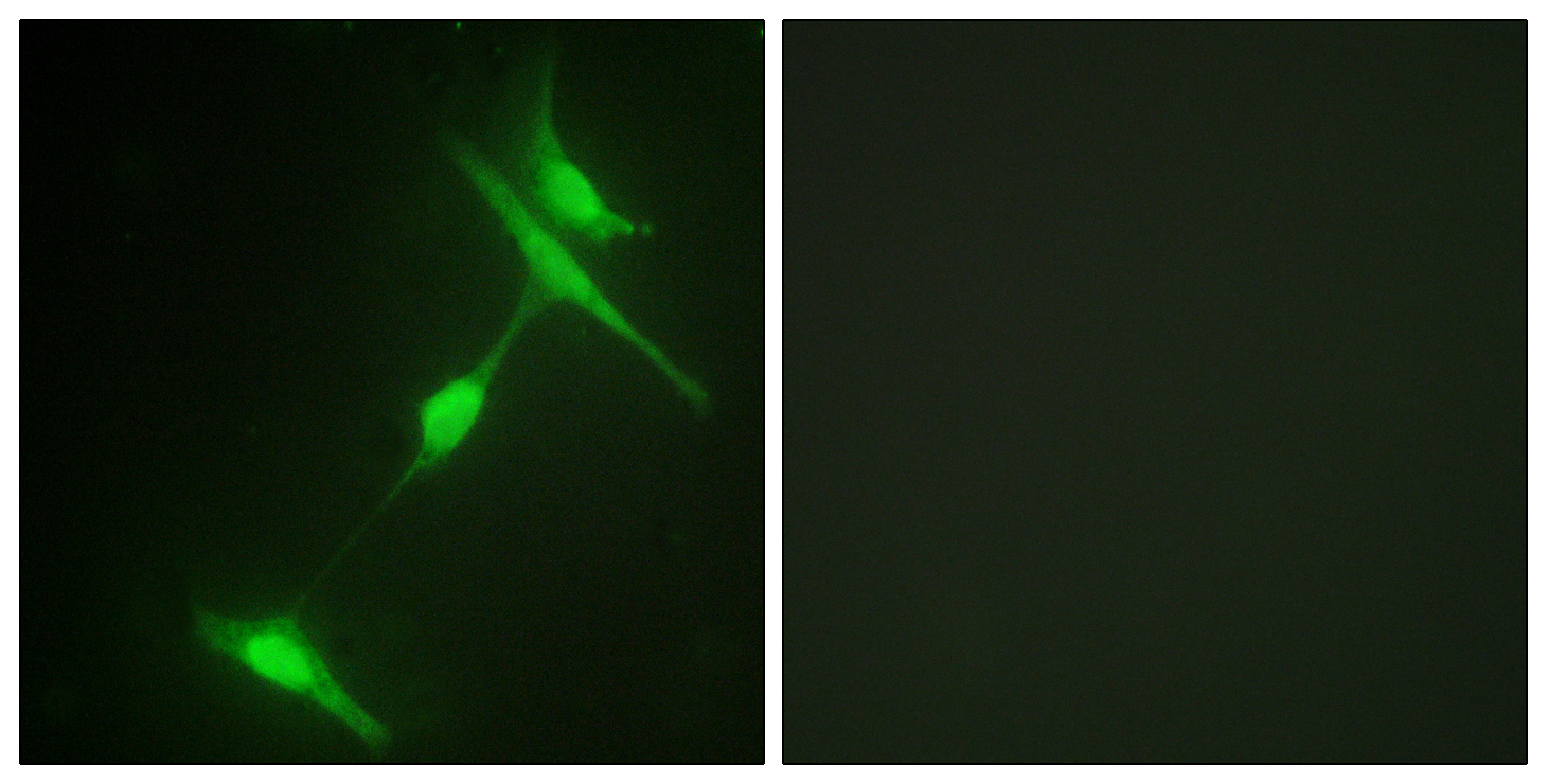
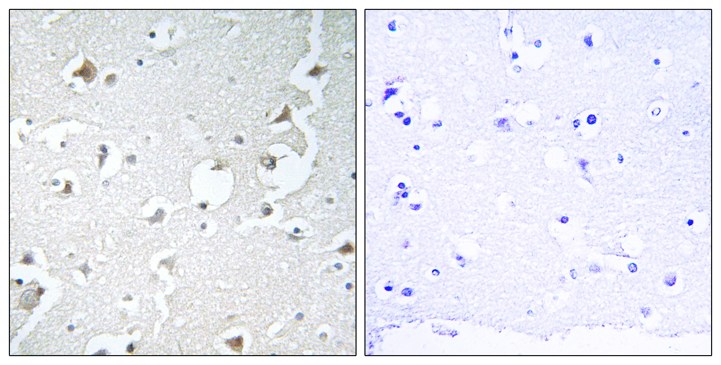
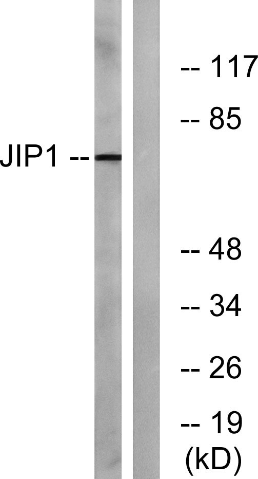
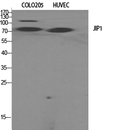
JIP-1 Polyclonal Antibody detects endogenous levels of JIP-1 protein.
宿主
Polyclonal, Rabbit,IgG
背景介绍
This gene encodes a regulator of the pancreatic beta-cell function. It is highly similar to JIP-1, a mouse protein known to be a regulator of c-Jun amino-terminal kinase (Mapk8). This protein has been shown to prevent MAPK8 mediated activation of transcription factors, and to decrease IL-1 beta and MAP kinase kinase 1 (MEKK1) induced apoptosis in pancreatic beta cells. This protein also functions as a DNA-binding transactivator of the glucose transporter GLUT2. RE1-silencing transcription factor (REST) is reported to repress the expression of this gene in insulin-secreting beta cells. This gene is found to be mutated in a type 2 diabetes family, and thus is thought to be a susceptibility gene for type 2 diabetes. [provided by RefSeq, May 2011],
组织表达
Highly expressed in brain. Expressed in neurons, localizing to neurite tips in differentiating cells. Also expressed in the pancreas, testis and prostate. Low levels in heart, ovary and small intestine. Decreased levels in pancreatic beta cells sensitize cells to IL-1-beta-induced apoptosis.
细胞定位
Cytoplasm . Cytoplasm, perinuclear region . Nucleus . Endoplasmic reticulum membrane. Mitochondrion membrane. Accumulates in cell surface projections. Under certain stress conditions, translocates to the perinuclear region of neurons. In insulin-secreting cells, detected in both the cytoplasm and nucleus (By similarity). .
信号通路
MAPK_ERK_Growth;MAPK_G_Protein;
功能
disease:Defects in MAPK8IP1 are a cause of non-insulin-dependent diabetes mellitus (NIDDM) [MIM:125853]. NIDDM is characterized by an autosomal dominant mode of inheritance, onset during adulthood and insulin resistance.,domain:A minimal inhibitory domain prevents pancreatic beta cell apoptosis in vitro, and prevents activation of c-jun by MAPK8, MAPK9 and MAPK10.,domain:The destruction boxes (D-box) may act as recognition signals for degradation via the ubiquitin-proteasome pathway.,function:The JNK-interacting protein (JIP) group of scaffold proteins selectively mediates JNK signaling by aggregating specific components of the MAPK cascade to form a functional JNK signaling module. Required for JNK activation in response to excitotoxic stress. Cytoplasmic MAPK8IP1 causes inhibition of JNK-regulated activity by retaining JNK in the cytoplasm and inhibiting JNK phosphorylation of c-Jun. May also participate in ApoER2-specific reelin signaling. Directly, or indirectly, regulates GLUT2 gene expression and beta-cell function. Appears to have a role in cell signaling in mature and developing nerve terminals. May function as a regulator of vesicle transport, through interactions with the JNK-signaling components and motor proteins (By similarity). Functions as an anti-apoptotic protein and whose level seems to influence the beta-cell death or survival response.,miscellaneous:A chemically synthesized cell-permeable peptide of the minimal inhibitory domain decreases brain lesions in both transient and permanent ischemia. The level of protection is still high when administered 6 or 12 hours after ischemia.,PTM:Phosphorylated by MAPK8, MAPK9 and MAPK10. Phosphorylation on Thr-103 is also necessary for the dissociation and activation of MAP3K12.,PTM:Ubiquitinated. Two preliminary events are required to prime for ubiquitination; phosphorylation and an increased in intracellular calcium concentration. Then, the calcium influx initiates ubiquitination and degradation by the ubiquitin-proteasome pathway.,similarity:Belongs to the JIP scaffold family.,similarity:Contains 1 PID domain.,similarity:Contains 1 SH3 domain.,subcellular location:Accumulates in cell surface projections. Under certain stress conditions, translocates to the perinuclear region of neurons. In insulin-secreting cells, detected in both the cytoplasm and nucleus.,subunit:Forms homo- or heterooligomeric complexes. Binds specific components of the JNK signaling pathway namely, MAPK8, MAPK9, MAPK10, MAPKK7, MLK2, MLK3, MAP3K12 and MAP3K13. Also binds the proline-rich domain-containing splice variant of apolipoprotein E receptor 2 (ApoER2). Interacts, via the PID domain, with RGNEF. Binds the cytoplasmic tails of LRP1 and LRP2 (Megalin). Binds the TPR motif-containing C-terminal of KNS2, then the pre-assembled MAPK8IP1 scaffolding complexes are transported as a cargo of kinesin, to the required subcellular location. Interacts with the cytoplasmic domain of APP.,tissue specificity:Highly expressed in brain. Expressed in neurons, localizing to neurite tips in differentiating cells. Also expressed in the pancreas, testis and prostate. Low levels in heart, ovary and small intestine. Decreased levels in pancreatic beta cells sensitize cells to IL-1-beta-induced apoptosis.,
纯化
The antibody was affinity-purified from rabbit antiserum by affinity-chromatography using epitope-specific immunogen.
.jpg)

