产品名称
IKKα Rabbit Polyclonal Antibody
别名
CHUK; IKKA; TCF16; Inhibitor of nuclear factor kappa-B kinase subunit alpha; I-kappa-B kinase alpha; IKK-A; IKK-alpha; IkBKA; IkappaB kinase; Conserved helix-loop-helix ubiquitous kinase; I-kappa-B kinase 1; IKK1; Nuclear factor NF-kappa-B
蛋白名称
Inhibitor of nuclear factor kappa-B kinase subunit alpha
存储缓冲液
Liquid in PBS containing 50% glycerol, 0.5% BSA and 0.02% New type preservative N.
Human Gene Link
http://www.ncbi.nlm.nih.gov/sites/entrez?db=gene&term=1147
Human Swissprot No.
O15111
Human Swissprot Link
http://www.uniprot.org/uniprotkb/O15111/entry
Mouse Swissprot No.
Q60680
Mouse Swissprot Link
http://www.uniprot.org/uniprot/Q60680
免疫原
The antiserum was produced against synthesized peptide derived from human IKK-alpha. AA range:15-64
特异性
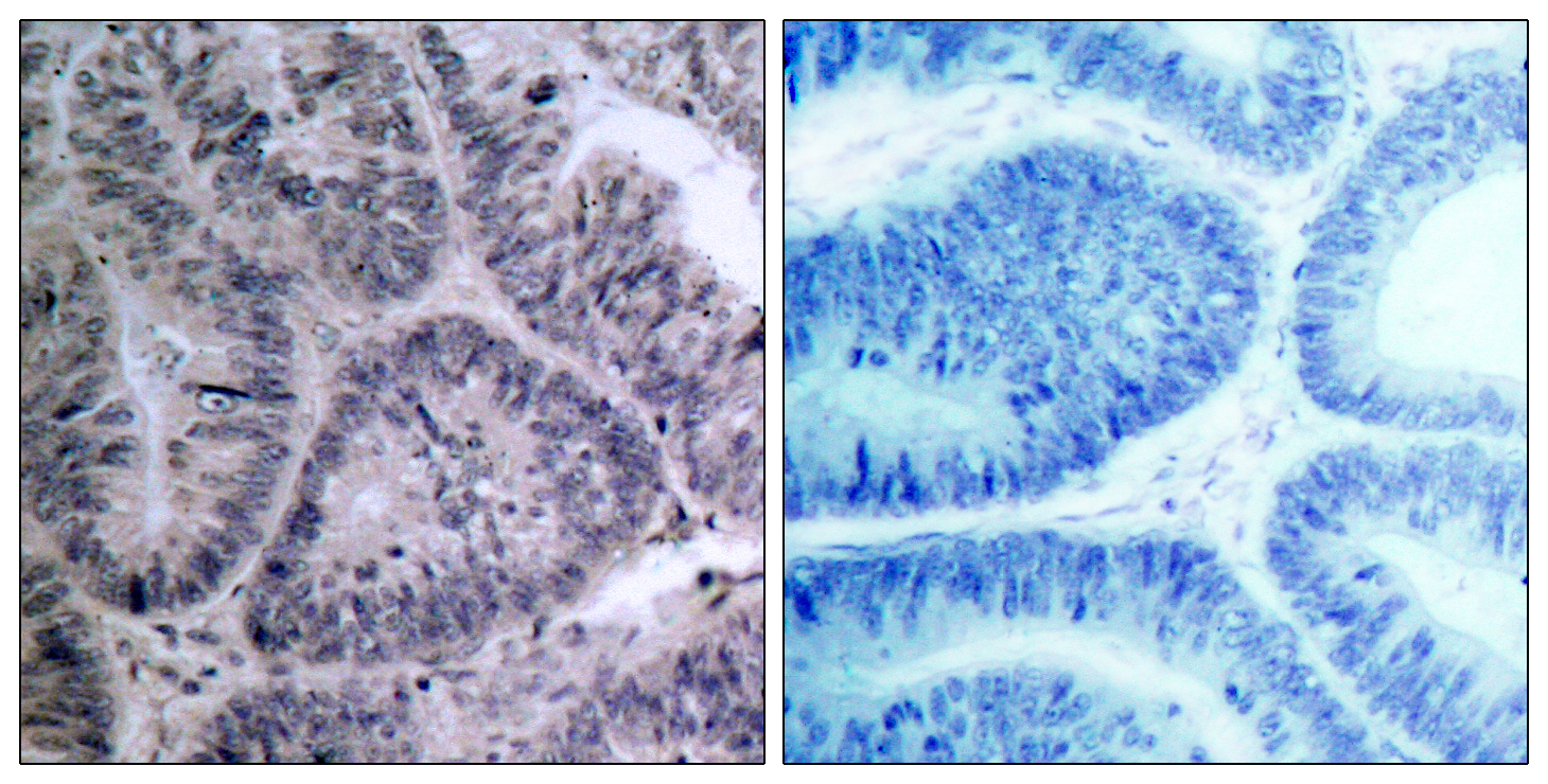
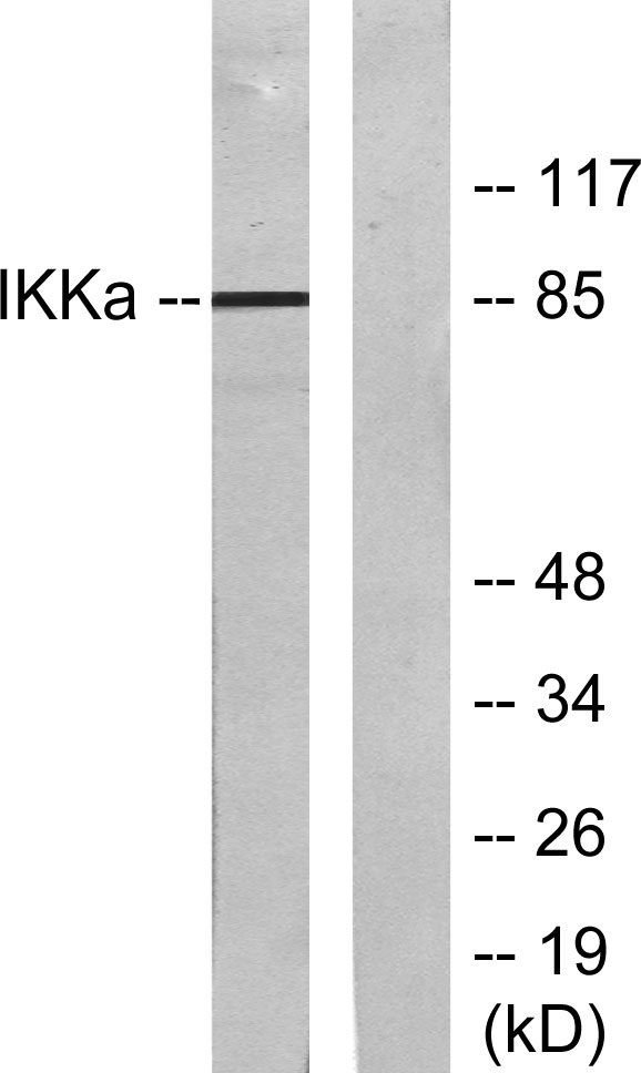
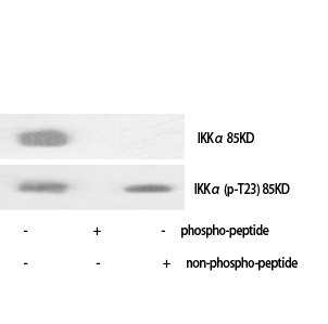
IKKα Polyclonal Antibody detects endogenous levels of IKKα protein.
宿主
Polyclonal, Rabbit,IgG
背景介绍
This gene encodes a member of the serine/threonine protein kinase family. The encoded protein, a component of a cytokine-activated protein complex that is an inhibitor of the essential transcription factor NF-kappa-B complex, phosphorylates sites that trigger the degradation of the inhibitor via the ubiquination pathway, thereby activating the transcription factor. [provided by RefSeq, Jul 2008],
细胞定位
Cytoplasm . Nucleus . Shuttles between the cytoplasm and the nucleus.
信号通路
T_Cell_Receptor; Insulin Receptor; B_Cell_Antigen; Stem cell pathway; Toll_Like; MAPK_ERK_Growth;MAPK_G_Protein; PI3K/Akt; NF_kappaB; Protein_Acetylation
功能
catalytic activity:ATP + [I-kappa-B protein] = ADP + [I-kappa-B phosphoprotein].,enzyme regulation:Activated when phosphorylated and inactivated when dephosphorylated.,function:Acts as part of the IKK complex in the conventional pathway of NF-kappa-B activation and phosphorylates inhibitors of NF-kappa-B thus leading to the dissociation of the inhibitor/NF-kappa-B complex and ultimately the degradation of the inhibitor. As part of the non-canonical pathway of NF-kappa-B activation, the MAP3K14-activated CHUK/IKKA homodimer phosphorylates NFKB2/p100 associated with RelB, inducing its proteolytic processing to NFKB2/p52 and the formation of NF-kappa-B RelB-p52 complexes. Also phosphorylates NCOA3. Phosphorylates 'Ser-10' of histone H3 at NF-kappa-B-regulated promoters during inflammatory responses triggered by cytokines.,PTM:Phosphorylated by MAP3K14/NIK, AKT and to a lesser extent by MEKK1, and dephosphorylated by PP2A. Autophosphorylated.,similarity:Belongs to the protein kinase superfamily.,similarity:Belongs to the protein kinase superfamily. Ser/Thr protein kinase family. I-kappa-B kinase subfamily.,similarity:Contains 1 protein kinase domain.,subcellular location:Shuttles between the cytoplasm and the nucleus.,subunit:Component of the I-kappa-B-kinase (IKK) core complex consisting of CHUK, IKBKB and IKBKG; probably four alpha/CHUK-beta/IKBKB dimers are associated with four gamma/IKBKG subunits. The IKK core complex seems to associate with regulatory or adapter proteins to form a IKK-signalosome holo-complex. Part of a complex composed of NCOA2, NCOA3, CHUK/IKKA, IKBKB, IKBKG and CREBBP. Part of a 70-90 kDa complex at least consisting of CHUK/IKKA, IKBKB, NFKBIA, RELA, IKBKAP and MAP3K14. Directly interacts with IKK-gamma/NEMO and TRPC4AP (By similarity). May interact with TRAF2. Interacts with NALP2. May interact with MAVS/IPS1.,tissue specificity:Widely expressed.,
纯化
The antibody was affinity-purified from rabbit antiserum by affinity-chromatography using epitope-specific immunogen.
.jpg)

