产品名称
ICAD Rabbit Polyclonal Antibody
别名
DFFA; DFF1; DFF45; H13; DNA fragmentation factor subunit alpha; DNA fragmentation factor 45 kDa subunit; DFF-45; Inhibitor of CAD; ICAD
蛋白名称
DNA fragmentation factor subunit alpha
存储缓冲液
Liquid in PBS containing 50% glycerol, 0.5% BSA and 0.02% New type preservative N.
Human Gene Link
http://www.ncbi.nlm.nih.gov/sites/entrez?db=gene&term=1676
Human Swissprot No.
O00273
Human Swissprot Link
http://www.uniprot.org/uniprotkb/O00273/entry
Mouse Gene Link
http://www.ncbi.nlm.nih.gov/sites/entrez?db=gene&term=13347
Mouse Swissprot No.
O54786
Mouse Swissprot Link
http://www.uniprot.org/uniprot/O54786
免疫原
The antiserum was produced against synthesized peptide derived from human DFFA. AA range:151-200
特异性
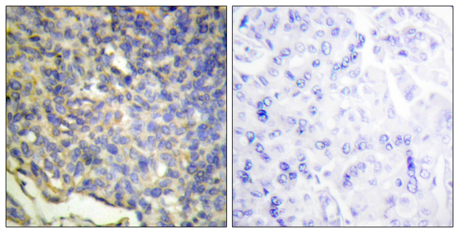
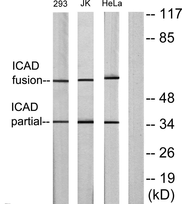
ICAD Polyclonal Antibody detects endogenous levels of ICAD protein.
宿主
Polyclonal, Rabbit,IgG
背景介绍
Apoptosis is a cell death process that removes toxic and/or useless cells during mammalian development. The apoptotic process is accompanied by shrinkage and fragmentation of the cells and nuclei and degradation of the chromosomal DNA into nucleosomal units. DNA fragmentation factor (DFF) is a heterodimeric protein of 40-kD (DFFB) and 45-kD (DFFA) subunits. DFFA is the substrate for caspase-3 and triggers DNA fragmentation during apoptosis. DFF becomes activated when DFFA is cleaved by caspase-3. The cleaved fragments of DFFA dissociate from DFFB, the active component of DFF. DFFB has been found to trigger both DNA fragmentation and chromatin condensation during apoptosis. Two alternatively spliced transcript variants encoding distinct isoforms have been found for this gene. [provided by RefSeq, Jul 2008],
组织表达
Breast,Coronary artery,Epithelium,Eye,Kidney,Skeletal muscle,
信号通路
Apoptosis_Inhibition;Apoptosis_Mitochondrial;Apoptosis_Overview;
功能
function:Inhibitor of the caspase-activated DNase (DFF40).,PTM:Caspase-3 cleaves DFF45 at 2 sites to generate an active factor.,PTM:Phosphorylated upon DNA damage, probably by ATM or ATR.,similarity:Contains 1 CIDE-N domain.,subunit:Heterodimer of DFFA and DFFB.,
纯化
The antibody was affinity-purified from rabbit antiserum by affinity-chromatography using epitope-specific immunogen.

.jpg)

