产品名称
Histone deacetylase 10 Rabbit Polyclonal Antibody
别名
HDAC10; Histone deacetylase 10; HD10
蛋白名称
Histone deacetylase 10
反应种属
Human,Mouse,Rat,Monkey
存储缓冲液
Liquid in PBS containing 50% glycerol, 0.5% BSA and 0.02% New type preservative N.
Human Gene Link
http://www.ncbi.nlm.nih.gov/sites/entrez?db=gene&term=83933
Human Swissprot No.
Q969S8
Human Swissprot Link
http://www.uniprot.org/uniprotkb/Q969S8/entry
Mouse Gene Link
http://www.ncbi.nlm.nih.gov/sites/entrez?db=gene&term=170787
Mouse Swissprot No.
Q6P3E7
Mouse Swissprot Link
http://www.uniprot.org/uniprot/Q6P3E7
Rat Gene Link
http://www.ncbi.nlm.nih.gov/sites/entrez?db=gene&term=362981
Rat Swissprot Link
http://www.uniprot.org/uniprot/Q569C4
免疫原
The antiserum was produced against synthesized peptide derived from human HDAC10. AA range:10-59
特异性
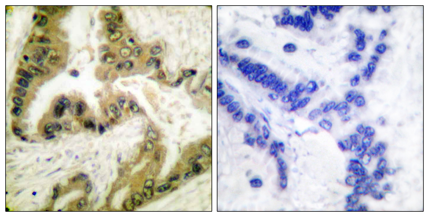
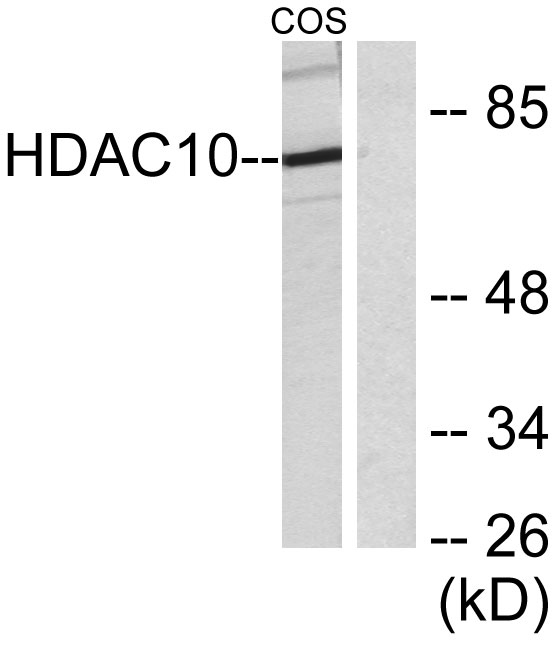
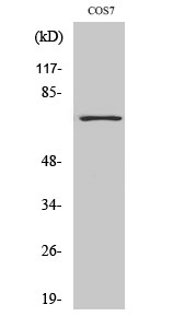
Histone deacetylase 10 Polyclonal Antibody detects endogenous levels of Histone deacetylase 10 protein.
宿主
Polyclonal, Rabbit,IgG
背景介绍
The protein encoded by this gene belongs to the histone deacetylase family, members of which deacetylate lysine residues on the N-terminal part of the core histones. Histone deacetylation modulates chromatin structure, and plays an important role in transcriptional regulation, cell cycle progression, and developmental events. Alternatively spliced transcript variants encoding different isoforms have been found for this gene. [provided by RefSeq, Aug 2011],
组织表达
Widely expressed with high levels in liver and kidney.
细胞定位
Cytoplasm . Nucleus . Excluded from nucleoli. .
功能
catalytic activity:Hydrolysis of an N(6)-acetyl-lysine residue of a histone to yield a deacetylated histone.,function:Responsible for the deacetylation of lysine residues on the N-terminal part of the core histones (H2A, H2B, H3 and H4). Histone deacetylation gives a tag for epigenetic repression and plays an important role in transcriptional regulation, cell cycle progression and developmental events. Histone deacetylases act via the formation of large multiprotein complexes.,similarity:Belongs to the histone deacetylase family. Type 2 subfamily.,subcellular location:Excluded from the nucleoli.,subunit:Interacts with HDAC2, HDAC3 and NCOR2.,tissue specificity:Ubiquitous. High expression in liver, spleen, pancreas and kidney.,
纯化
The antibody was affinity-purified from rabbit antiserum by affinity-chromatography using epitope-specific immunogen.