产品名称
Golgin 45 Rabbit Polyclonal Antibody
别名
BLZF1; JEM1; Golgin-45; Basic leucine zipper nuclear factor 1; JEM-1; p45 basic leucine-zipper nuclear factor
存储缓冲液
Liquid in PBS containing 50% glycerol, 0.5% BSA and 0.02% New type preservative N.
Human Gene Link
http://www.ncbi.nlm.nih.gov/sites/entrez?db=gene&term=8548
Human Swissprot No.
Q9H2G9
Human Swissprot Link
http://www.uniprot.org/uniprotkb/Q9H2G9/entry
Mouse Gene Link
http://www.ncbi.nlm.nih.gov/sites/entrez?db=gene&term=66352
Mouse Swissprot No.
Q8R2X8
Mouse Swissprot Link
http://www.uniprot.org/uniprot/Q8R2X8
免疫原
The antiserum was produced against synthesized peptide derived from human BLZF1. AA range:10-59
特异性
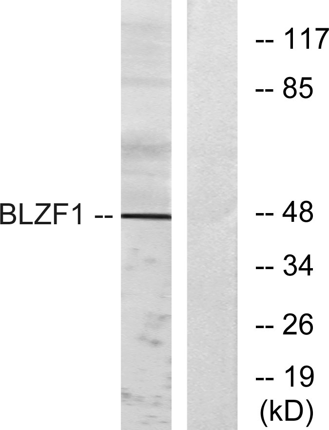
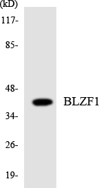
Golgin 45 Polyclonal Antibody detects endogenous levels of Golgin 45 protein.
宿主
Polyclonal, Rabbit,IgG
背景介绍
caution:Because of the presence of a potential basic motif and leucine-zipper domain, PubMed:9129147 and PubMed:11056056 have thought that BLZF1 is a potential transcription factor. They found it localized in the nucleus, except isoform 2, which was cytoplasmic. However, homology at several typical position for basic or hydrophobic residues is missing.,function:Required for normal Golgi structure and for protein transport from the endoplasmic reticulum (ER) through the Golgi apparatus to the cell surface.,induction:Up-regulated by retinoids.,subunit:Interacts with GORASP2 and with the GTP-bound form of RAB2, but not with other Golgi Rab proteins. GORASP2 and BLZF1 form a RAB2 effector complex on medial Golgi.,tissue specificity:Ubiquitous. Also found in cell lines derived from several hematopoietic pathologies, such as T-cell leukemia, pro-B, pre-B, myeloma, and plasmacytoma cell lines, but not in Burkitt lymphoma cells.,
组织表达
Detected in adrenal gland (PubMed:9129147).
细胞定位
Golgi apparatus membrane .; [Isoform 1]: Nucleus . Detected in the nucleus upon heterologous expression. Not detected in the cytoplasm. .; [Isoform 2]: Cytoplasm . Not detected in the nucleus. .
功能
caution:Because of the presence of a potential basic motif and leucine-zipper domain, PubMed:9129147 and PubMed:11056056 have thought that BLZF1 is a potential transcription factor. They found it localized in the nucleus, except isoform 2, which was cytoplasmic. However, homology at several typical position for basic or hydrophobic residues is missing.,function:Required for normal Golgi structure and for protein transport from the endoplasmic reticulum (ER) through the Golgi apparatus to the cell surface.,induction:Up-regulated by retinoids.,subunit:Interacts with GORASP2 and with the GTP-bound form of RAB2, but not with other Golgi Rab proteins. GORASP2 and BLZF1 form a RAB2 effector complex on medial Golgi.,tissue specificity:Ubiquitous. Also found in cell lines derived from several hematopoietic pathologies, such as T-cell leukemia, pro-B, pre-B, myeloma, and plasmacytoma cell lines, but not in Burkitt lymphoma cells.,
纯化
The antibody was affinity-purified from rabbit antiserum by affinity-chromatography using epitope-specific immunogen.