FEN-1 Rabbit Polyclonal Antibody
产品基本信息
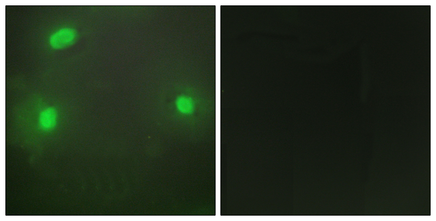
Immunofluorescence analysis of HeLa cells, using FEN1 Antibody. The picture on the right is blocked with the synthesized peptide.
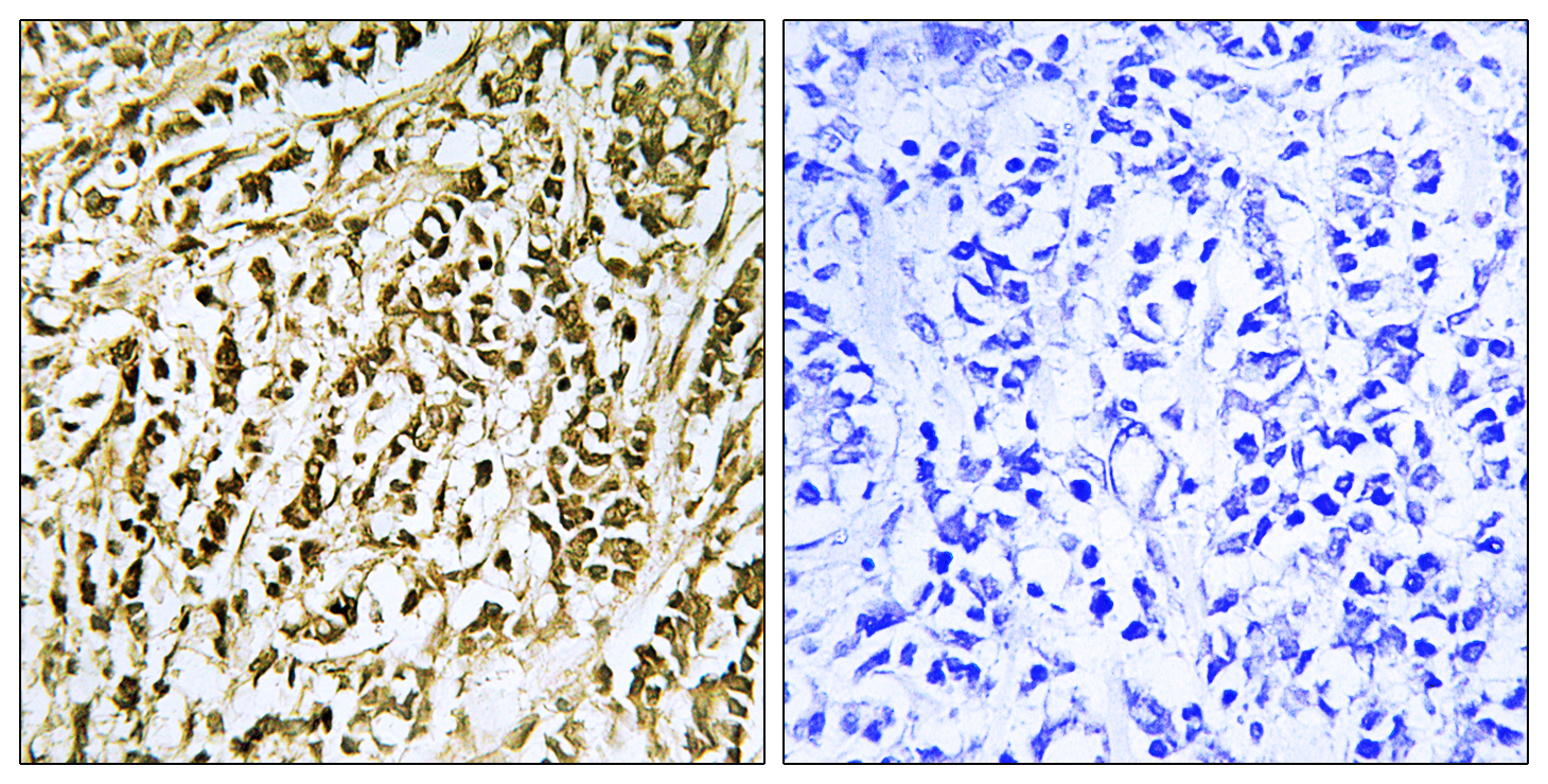
Immunohistochemistry analysis of paraffin-embedded human breast carcinoma tissue, using FEN1 Antibody. The picture on the right is blocked with the synthesized peptide.
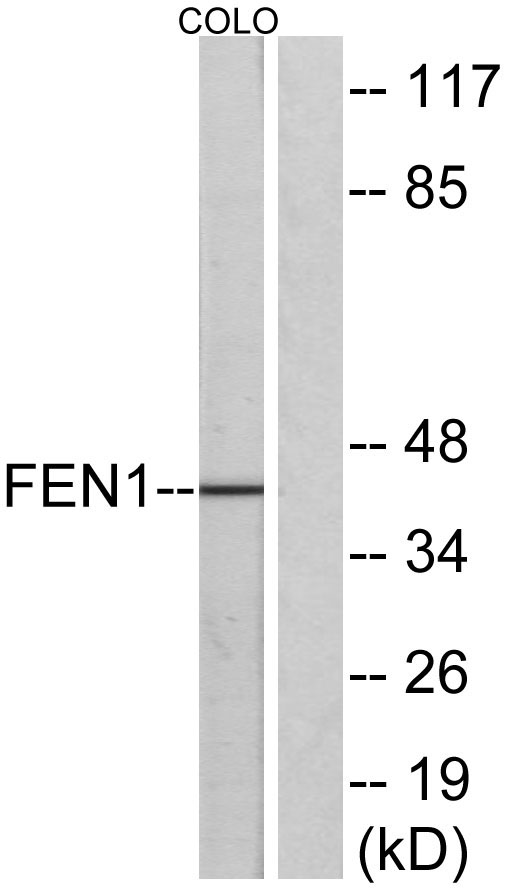
Western blot analysis of lysates from COLO205 cells, using FEN1 Antibody. The lane on the right is blocked with the synthesized peptide.
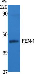
Western Blot analysis of various cells using FEN-1 Polyclonal Antibody diluted at 1:500
.jpg)
Western Blot analysis of HuvEc cells using FEN-1 Polyclonal Antibody diluted at 1:500
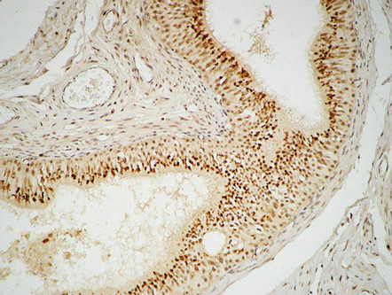
Immunohistochemical analysis of paraffin-embedded Human testis. 1, Antibody was diluted at 1:100(4°,overnight). 2, High-pressure and temperature EDTA, pH8.0 was used for antigen retrieval. 3,Secondary antibody was diluted at 1:200(room temperature, 30min).
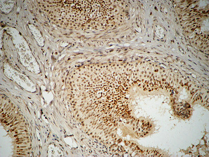
Immunohistochemical analysis of paraffin-embedded Human testis. 1, Antibody was diluted at 1:100(4°,overnight). 2, High-pressure and temperature EDTA, pH8.0 was used for antigen retrieval. 3,Secondary antibody was diluted at 1:200(room temperature, 30min).
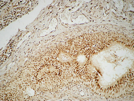
Immunohistochemical analysis of paraffin-embedded Human testis. 1, Antibody was diluted at 1:100(4°,overnight). 2, High-pressure and temperature EDTA, pH8.0 was used for antigen retrieval. 3,Secondary antibody was diluted at 1:200(room temperature, 30min).
相关文献
产品问答
相关产品

市场:027-65023363 行政/人事:027-62439686 邮箱:marketing@brainvta.com 客服:18140661572(活动咨询、售后反馈等)
销售总监:张经理 18995532642 华东区:陈经理 18013970337 华南区:王经理 13100653525 华中/西区:杨经理 18186518905 华北区:张经理 18893721749
地址:中国武汉东湖高新区光谷七路128号中科开物产业园1号楼
Copyright © 武汉枢密脑科学技术有限公司. All RIGHTS RESERVED.
鄂ICP备2021009124号 DIGITAL BY VTHINK
