产品名称
Fe65L Rabbit Polyclonal Antibody
别名
APBB2; FE65L; FE65L1; Amyloid beta A4 precursor protein-binding family B member 2; Protein Fe65-like 1
蛋白名称
Amyloid beta A4 precursor protein-binding family B member 2
存储缓冲液
Liquid in PBS containing 50% glycerol, 0.5% BSA and 0.02% New type preservative N.
Human Gene Link
http://www.ncbi.nlm.nih.gov/sites/entrez?db=gene&term=323
Human Swissprot No.
Q92870
Human Swissprot Link
http://www.uniprot.org/uniprotkb/Q92870/entry
Mouse Gene Link
http://www.ncbi.nlm.nih.gov/sites/entrez?db=gene&term=11787
Mouse Swissprot No.
Q9DBR4
Mouse Swissprot Link
http://www.uniprot.org/uniprot/Q9DBR4
免疫原
The antiserum was produced against synthesized peptide derived from human APBB2. AA range:471-520
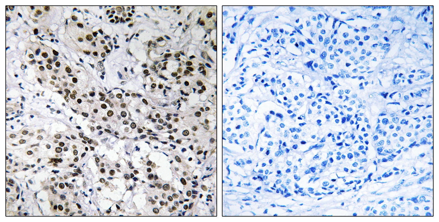
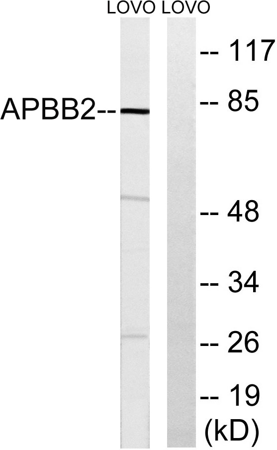
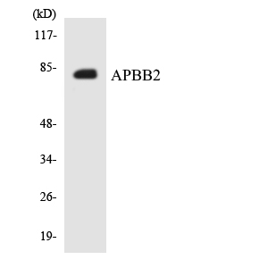
特异性
Fe65L Polyclonal Antibody detects endogenous levels of Fe65L protein.
宿主
Polyclonal, Rabbit,IgG
背景介绍
amyloid beta precursor protein binding family B member 2(APBB2) Homo sapiens The protein encoded by this gene interacts with the cytoplasmic domains of amyloid beta (A4) precursor protein and amyloid beta (A4) precursor-like protein 2. This protein contains two phosphotyrosine binding (PTB) domains, which are thought to function in signal transduction. Polymorphisms in this gene have been associated with Alzheimer's disease. Alternative splicing results in multiple transcript variants. [provided by RefSeq, Oct 2009],
细胞定位
Endoplasmic reticulum . Golgi apparatus . Early endosome .
功能
function:May modulate the internalization of beta-amyloid precursor protein.,similarity:Contains 1 WW domain.,similarity:Contains 2 PID domains.,subunit:Binds to the intracellular domain of the beta-amyloid precursor protein.,
纯化
The antibody was affinity-purified from rabbit antiserum by affinity-chromatography using epitope-specific immunogen.