Cn|En

现货抗体产品库

FAK Rabbit Polyclonal Antibody

产品详情 相关文献 产品问答 相关产品

产品基本信息

产品货号
BD-PT1659
产品名称
FAK Rabbit Polyclonal Antibody
别名
PTK2; FAK; FAK1; Focal adhesion kinase 1; FADK 1; Focal adhesion kinase-related nonkinase; FRNK; Protein phosphatase 1 regulatory subunit 71; PPP1R71; Protein-tyrosine kinase 2; p125FAK; pp125FAK
类别
常规抗体
基因名称
PTK2
蛋白名称
Focal adhesion kinase 1
推荐应用
WB
反应种属
Human,Mouse,Rat
浓度
1 mg/ml
存储缓冲液
Liquid in PBS containing 50% glycerol, 0.5% BSA and 0.02% New type preservative N.
Human Gene ID
5747
Human Gene Link
http://www.ncbi.nlm.nih.gov/sites/entrez?db=gene&term=5747
Human Swissprot No.
Q05397
Human Swissprot Link
http://www.uniprot.org/uniprotkb/Q05397/entry
Mouse Gene ID
14083
Mouse Gene Link
http://www.ncbi.nlm.nih.gov/sites/entrez?db=gene&term=14083
Mouse Swissprot No.
P34152
Mouse Swissprot Link
http://www.uniprot.org/uniprot/P34152
Rat Gene ID
25614
Rat Gene Link
http://www.ncbi.nlm.nih.gov/sites/entrez?db=gene&term=25614
Rat Swissprot No.
O35346
Rat Swissprot Link
http://www.uniprot.org/uniprot/O35346
免疫原
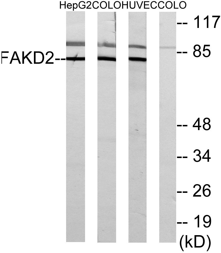
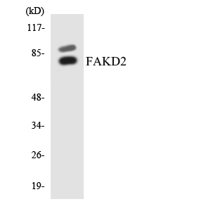
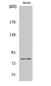
The antiserum was produced against synthesized peptide derived from human FAK. AA range:809-858
特异性
FAK Polyclonal Antibody detects endogenous levels of FAK protein.
稀释度
IF 1:50-200 WB 1:500 - 1:2000. IHC 1:100 - 1:300. ELISA: 1:40000. Not yet tested in other applications.
预测分子量
120kD
运输及保存条件
-20°C/1 year
宿主
Polyclonal, Rabbit,IgG
背景介绍
protein tyrosine kinase 2(PTK2) Homo sapiens This gene encodes a cytoplasmic protein tyrosine kinase which is found concentrated in the focal adhesions that form between cells growing in the presence of extracellular matrix constituents. The encoded protein is a member of the FAK subfamily of protein tyrosine kinases but lacks significant sequence similarity to kinases from other subfamilies. Activation of this gene may be an important early step in cell growth and intracellular signal transduction pathways triggered in response to certain neural peptides or to cell interactions with the extracellular matrix. Several transcript variants encoding different isoforms have been found for this gene, but the full-length natures of only four of them have been determined. [provided by RefSeq, Oct 2015],
组织表达
Detected in B and T-lymphocytes. Isoform 1 and isoform 6 are detected in lung fibroblasts (at protein level). Ubiquitous. Expressed in epithelial cells (at protein level) (PubMed:31630787).
细胞定位
Cell junction, focal adhesion. Cell membrane; Peripheral membrane protein; Cytoplasmic side. Cytoplasm, cell cortex. Cytoplasm, cytoskeleton. Cytoplasm, cytoskeleton, microtubule organizing center, centrosome . Nucleus. Cytoplasm, cytoskeleton, cilium basal body . Constituent of focal adhesions. Detected at microtubules.
信号通路
ErbB_HER;Chemokine;Axon guidance;VEGF;Focal adhesion;Leukocyte transendothelial migration;Regulates Actin and Cytoskeleton;Pathways in cancer;Small cell lung cancer;
功能
catalytic activity:ATP + a [protein]-L-tyrosine = ADP + a [protein]-L-tyrosine phosphate.,domain:The carboxy-terminal region is the site of focal adhesion targeting (FAT) sequence which mediates the localization of FAK1 to focal adhesions.,domain:The first Pro-rich domain interacts with the SH3 domain of CRK-associated substrate (BCAR1) and CASL.,function:Non-receptor protein-tyrosine kinase implicated in signaling pathways involved in cell motility, proliferation and apoptosis. Activated by tyrosine-phosphorylation in response to either integrin clustering induced by cell adhesion or antibody cross-linking, or via G-protein coupled receptor (GPCR) occupancy by ligands such as bombesin or lysophosphatidic acid, or via LDL receptor occupancy. Plays a potential role in oncogenic transformations resulting in increased kinase activity.,PTM:Phosphorylated on 6 tyrosine residues upon activation.,similarity:Belongs to the protein kinase superfamily. Tyr protein kinase family.,similarity:Belongs to the protein kinase superfamily. Tyr protein kinase family. FAK subfamily.,similarity:Contains 1 FERM domain.,similarity:Contains 1 protein kinase domain.,subcellular location:Constituent of focal adhesions.,subunit:Interacts with CAS family members and with GIT1, SORBS1 and BCAR3. Interacts with RGNEF and SHB (By similarity). Interacts with TGFB1I1.,tissue specificity:Expressed in all organs tested, in lymphoid cell lines, but most abundantly in brain.,
期货
现货
纯化
The antibody was affinity-purified from rabbit antiserum by affinity-chromatography using epitope-specific immunogen.

Immunohistochemistry analysis of paraffin-embedded human colon carcinoma tissue, using FAK Antibody. The picture on the right is blocked with the synthesized peptide.

Western blot analysis of lysates from MCF-7 and HUVEC cells, using FAK Antibody. The lane on the right is blocked with the synthesized peptide.

Western blot analysis of the lysates from K562 cells using FAK antibody.

Immunofluorescence analysis of human-liver tissue. 1,FAK Polyclonal Antibody(red) was diluted at 1:200(4°C,overnight). 2, Cy3 labled Secondary antibody was diluted at 1:300(room temperature, 50min).3, Picture B: DAPI(blue) 10min. Picture A:Target. Picture B: DAPI. Picture C: merge of A+B

Immunofluorescence analysis of human-liver tissue. 1,FAK Polyclonal Antibody(red) was diluted at 1:200(4°C,overnight). 2, Cy3 labled Secondary antibody was diluted at 1:300(room temperature, 50min).3, Picture B: DAPI(blue) 10min. Picture A:Target. Picture B: DAPI. Picture C: merge of A+B

Immunofluorescence analysis of human-lung tissue. 1,FAK Polyclonal Antibody(red) was diluted at 1:200(4°C,overnight). 2, Cy3 labled Secondary antibody was diluted at 1:300(room temperature, 50min).3, Picture B: DAPI(blue) 10min. Picture A:Target. Picture B: DAPI. Picture C: merge of A+B

Immunofluorescence analysis of human-lung tissue. 1,FAK Polyclonal Antibody(red) was diluted at 1:200(4°C,overnight). 2, Cy3 labled Secondary antibody was diluted at 1:300(room temperature, 50min).3, Picture B: DAPI(blue) 10min. Picture A:Target. Picture B: DAPI. Picture C: merge of A+B

Immunofluorescence analysis of rat-kidney tissue. 1,FAK Polyclonal Antibody(red) was diluted at 1:200(4°C,overnight). 2, Cy3 labled Secondary antibody was diluted at 1:300(room temperature, 50min).3, Picture B: DAPI(blue) 10min. Picture A:Target. Picture B: DAPI. Picture C: merge of A+B

Immunofluorescence analysis of rat-kidney tissue. 1,FAK Polyclonal Antibody(red) was diluted at 1:200(4°C,overnight). 2, Cy3 labled Secondary antibody was diluted at 1:300(room temperature, 50min).3, Picture B: DAPI(blue) 10min. Picture A:Target. Picture B: DAPI. Picture C: merge of A+B

Western Blot analysis of various cells using primary antibody diluted at 1:1000(4°C overnight). Secondary antibody:Goat Anti-rabbit IgG IRDye 800( diluted at 1:5000, 25°C, 1 hour). Cell lysate was extracted by Minute™ Plasma Membrane Protein Isolation and Cell Fractionation Kit(SM-005, Inventbiotech,MN,USA).

Western blot analysis of HELA KB SH-SY5Y 293T 3T3 lysis using FAK antibody. Antibody was diluted at 1:2000

Western blot analysis of customer‘s lysis using FAK antibody. Antibody was diluted at 1:2000

相关文献

产品问答

相关产品

免责声明| 法律支持| 联系方式

市场:027-65023363   行政/人事:027-62439686   邮箱:marketing@brainvta.com  

销售总监:张经理  18995532642  华东区:陈经理 18013970337   华南区:王经理 13100653525   华中/西区:杨经理 18186518905   华北区:张经理 18893721749

地址:中国武汉东湖高新区光谷七路128号中科开物产业园1号楼

Copyright © 武汉枢密脑科学技术有限公司. All RIGHTS RESERVED.
鄂ICP备2021009124号 DIGITAL BY VTHINK