Cn|En

现货抗体产品库

Ezrin Rabbit Polyclonal Antibody

产品详情 相关文献 产品问答 相关产品

产品基本信息

产品货号
BD-PT1651
产品名称
Ezrin Rabbit Polyclonal Antibody
别名
EZR; VIL2; Ezrin; Cytovillin; Villin-2; p81
类别
常规抗体
基因名称
EZR
蛋白名称
Ezrin
推荐应用
WB
反应种属
Human,Mouse,Rat
浓度
1 mg/ml
存储缓冲液
Liquid in PBS containing 50% glycerol, 0.5% BSA and 0.02% New type preservative N.
Human Gene ID
7430
Human Gene Link
http://www.ncbi.nlm.nih.gov/sites/entrez?db=gene&term=7430
Human Swissprot No.
P15311
Human Swissprot Link
http://www.uniprot.org/uniprotkb/P15311/entry
Mouse Gene ID
22350
Mouse Gene Link
http://www.ncbi.nlm.nih.gov/sites/entrez?db=gene&term=22350
Mouse Swissprot No.
P26040
Mouse Swissprot Link
http://www.uniprot.org/uniprot/P26040
Rat Gene ID
54319
Rat Gene Link
http://www.ncbi.nlm.nih.gov/sites/entrez?db=gene&term=54319
Rat Swissprot No.
P31977
Rat Swissprot Link
http://www.uniprot.org/uniprot/P31977
免疫原
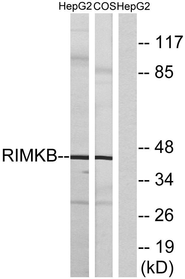
The antiserum was produced against synthesized peptide derived from human Ezrin. AA range:511-560
特异性
Ezrin Polyclonal Antibody detects endogenous levels of Ezrin protein.
稀释度
WB 1:500 - 1:2000. ELISA: 1:20000.. IF 1:50-200
预测分子量
70kD
运输及保存条件
-20°C/1 year
宿主
Polyclonal, Rabbit,IgG
背景介绍
The cytoplasmic peripheral membrane protein encoded by this gene functions as a protein-tyrosine kinase substrate in microvilli. As a member of the ERM protein family, this protein serves as an intermediate between the plasma membrane and the actin cytoskeleton. This protein plays a key role in cell surface structure adhesion, migration and organization, and it has been implicated in various human cancers. A pseudogene located on chromosome 3 has been identified for this gene. Alternatively spliced variants have also been described for this gene. [provided by RefSeq, Jul 2008],
组织表达
Expressed in cerebral cortex, basal ganglia, hippocampus, hypophysis, and optic nerve. Weakly expressed in brain stem and diencephalon. Stronger expression was detected in gray matter of frontal lobe compared to white matter (at protein level). Component of the microvilli of intestinal epithelial cells. Preferentially expressed in astrocytes of hippocampus, frontal cortex, thalamus, parahippocampal cortex, amygdala, insula, and corpus callosum. Not detected in neurons in most tissues studied.
细胞定位
Apical cell membrane ; Peripheral membrane protein ; Cytoplasmic side . Cell projection . Cell projection, microvillus membrane ; Peripheral membrane protein ; Cytoplasmic side . Cell projection, ruffle membrane ; Peripheral membrane protein ; Cytoplasmic side . Cytoplasm, cell cortex . Cytoplasm, cytoskeleton . Cell projection, microvillus . Localization to the apical membrane of parietal cells depends on the interaction with PALS1. Localizes to cell extensions and peripheral processes of astrocytes (By similarity). Microvillar peripheral membrane protein (cytoplasmic side). .
信号通路
Leukocyte transendothelial migration;Regulates Actin and Cytoskeleton;Pathogenic Escherichia coli infection;
功能
developmental stage:Very strong staining is detected in the Purkinje cell layer and in part of the molecular layer of the infant brain compared to adult brain.,function:Probably involved in connections of major cytoskeletal structures to the plasma membrane. In epithelial cells, required for the formation of microvilli and membrane ruffles on the apical pole. Along with PLEKHG6, required for normal macropinocytosis.,PTM:Phosphorylated by tyrosine-protein kinases.,similarity:Contains 1 FERM domain.,subcellular location:Localization to the apical membrane of parietal cells depends on the interaction with MPP5. Localizes to cell extensions and peripheral processes of astrocytes (By similarity). Microvillar peripheral membrane protein (cytoplasmic side).,subunit:Interacts with MPP5 (By similarity). Interacts with SLC9A3R1 and SCYL3/PACE1. Interacts with PLEKHG6. Interacts with NGX6.,tissue specificity:Expressed in cerebral cortex, basal ganglia, hippocampus, hypophysis, and optic nerve. Weakly expressed in brain stem and diencephalon. Stronger expression was detected in gray matter of frontal lobe compared to white matter (at protein level). Component of the microvilli of intestinal epithelial cells. Preferentially expressed in astrocytes of hippocampus, frontal cortex, thalamus, parahippocampal cortex, amygdala, insula, and corpus callosum. Not detected in neurons in most tissues studied.,
期货
现货
纯化
The antibody was affinity-purified from rabbit antiserum by affinity-chromatography using epitope-specific immunogen.

Immunohistochemistry analysis of paraffin-embedded human brain tissue, using Ezrin Antibody. The picture on the right is blocked with the synthesized peptide.

Western blot analysis of lysates from HuvE, MCF-7, HepG2, and HeLa cells, using Ezrin Antibody. The lane on the right is blocked with the synthesized peptide.

Western Blot analysis of various cells using Ezrin Polyclonal Antibody diluted at 1:2000

Western Blot analysis of MCF7 cells using Ezrin Polyclonal Antibody diluted at 1:2000

相关文献

产品问答

相关产品

免责声明| 法律支持| 联系方式

市场:027-65023363   行政/人事:027-62439686   邮箱:marketing@brainvta.com  

销售总监:张经理  18995532642  华东区:陈经理 18013970337   华南区:王经理 13100653525   华中/西区:杨经理 18186518905   华北区:张经理 18893721749

地址:中国武汉东湖高新区光谷七路128号中科开物产业园1号楼

Copyright © 武汉枢密脑科学技术有限公司. All RIGHTS RESERVED.
鄂ICP备2021009124号 DIGITAL BY VTHINK