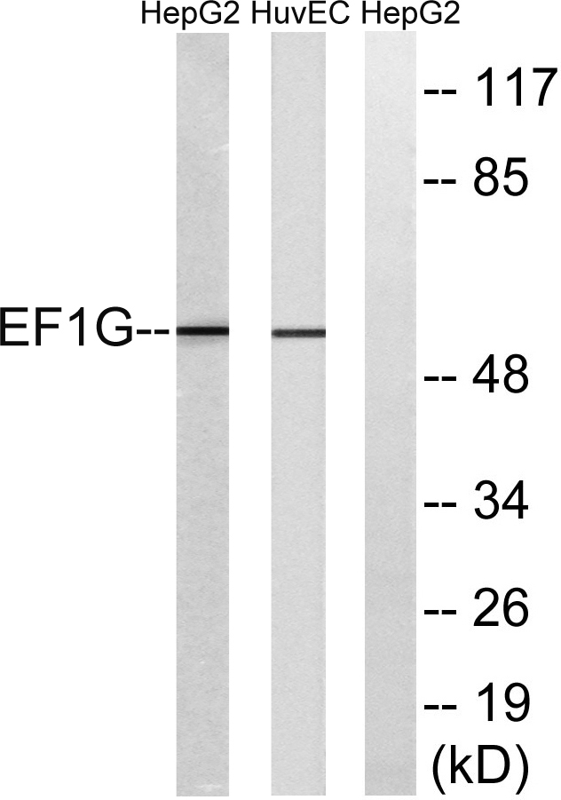
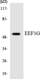
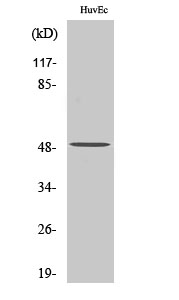
Cn|En

现货抗体产品库

E-cadherin Rabbit Polyclonal Antibody

产品详情 相关文献 产品问答 相关产品

产品基本信息

产品货号
BD-PT1454
产品名称
E-cadherin Rabbit Polyclonal Antibody
别名
CDH1; CDHE; UVO; Cadherin-1; CAM 120/80; Epithelial cadherin; E-cadherin; Uvomorulin; CD antigen CD324; CDH2; CDHN; NCAD; Cadherin-2; CDw325; Neural cadherin; N-cadherin; CD antigen CD325; CDH3; CDHP; Cadherin-3; Placental cadherin; P-cadhe
类别
常规抗体
基因名称
CDH1
蛋白名称
Cadherin-1
推荐应用
WB
反应种属
Human,Mouse,Rat
浓度
1 mg/ml
存储缓冲液
Liquid in PBS containing 50% glycerol, 0.5% BSA and 0.02% New type preservative N.
Human Gene ID
999/1000/1001/1002
Human Gene Link
http://www.ncbi.nlm.nih.gov/sites/entrez?db=gene&term=999
Human Swissprot No.
P12830/P19022/P22223/P55283
Human Swissprot Link
http://www.uniprot.org/uniprotkb/P12830/entry
Mouse Gene ID
12550/12558/12560/12561
Mouse Gene Link
http://www.ncbi.nlm.nih.gov/sites/entrez?db=gene&term=12550
Rat Gene ID
83502/83501
Rat Gene Link
http://www.ncbi.nlm.nih.gov/sites/entrez?db=gene&term=83502
Rat Swissprot No.
Q9R0T4/Q9Z1Y3/Q63149
Rat Swissprot Link
http://www.uniprot.org/uniprot/Q9R0T4
免疫原
The antiserum was produced against synthesized peptide derived from human Cadherin. AA range:833-882
特异性
E-cadherin Polyclonal Antibody detects endogenous levels of E-cadherin protein.
稀释度
WB 1:500 - 1:2000. IHC-p: 1:100-300 ELISA: 1:20000. IF 1:100-300 Not yet tested in other applications.
预测分子量
125-130kD
运输及保存条件
-20°C/1 year
宿主
Polyclonal, Rabbit,IgG
背景介绍
This gene encodes a classical cadherin of the cadherin superfamily. Alternative splicing results in multiple transcript variants, at least one of which encodes a preproprotein that is proteolytically processed to generate the mature glycoprotein. This calcium-dependent cell-cell adhesion protein is comprised of five extracellular cadherin repeats, a transmembrane region and a highly conserved cytoplasmic tail. Mutations in this gene are correlated with gastric, breast, colorectal, thyroid and ovarian cancer. Loss of function of this gene is thought to contribute to cancer progression by increasing proliferation, invasion, and/or metastasis. The ectodomain of this protein mediates bacterial adhesion to mammalian cells and the cytoplasmic domain is required for internalization. This gene is present in a gene cluster with other members of the cadherin family on chromosome 16. [provided by RefSeq, Nov 2015],
组织表达
Non-neural epithelial tissues.
细胞定位
Cell junction, adherens junction . Cell membrane ; Single-pass type I membrane protein. Endosome. Golgi apparatus, trans-Golgi network. Colocalizes with DLGAP5 at sites of cell-cell contact in intestinal epithelial cells. Anchored to actin microfilaments through association with alpha-, beta- and gamma-catenin. Sequential proteolysis induced by apoptosis or calcium influx, results in translocation from sites of cell-cell contact to the cytoplasm. Colocalizes with RAB11A endosomes during its transport from the Golgi apparatus to the plasma membrane.
信号通路
Cell adhesion molecules (CAMs);Adherens_Junction;Pathogenic Escherichia coli infection;Pathways in cancer;Endometrial cancer;Thyroid cancer;Melanoma;Bladder cancer;
功能
disease:Defects in CDH1 are a cause of gastric cancer [MIM:137215]; also known as hereditary familial diffuse gastric cancer (HDGC).,disease:Defects in CDH1 are a cause of susceptibility to endometrial cancer [MIM:608089].,disease:Defects in CDH1 are associated with ovarian cancer [MIM:167000]. Ovarian cancer is the leading cause of death from gynecologic malignancy. It is characterized by advanced presentation with loco-regional dissemination in the peritoneal cavity and the rare incidence of visceral metastases. These typical features relate to the biology of the disease, which is a principal determinant of outcome.,disease:Defects in CDH1 are involved in dysfunction of the cell-cell adhesion system, triggering cancer invasion (gastric, breast, ovary, endometrium and thyroid) and metastasis.,function:Cadherins are calcium dependent cell adhesion proteins.,function:Cadherins are calcium-dependent cell adhesion proteins. They preferentially interact with themselves in a homophilic manner in connecting cells; cadherins may thus contribute to the sorting of heterogeneous cell types. CDH1 is involved in mechanisms regulating cell-cell adhesions, mobility and proliferation of epithelial cells. Has a potent invasive suppressor role. It is a ligand for integrin alpha-E/beta-7.,function:E-Cad/CTF2 promotes non-amyloidogenic degradation of Abeta precursors. Has a strong inhibitory effect on APP C99 and C83 production.,online information:E-cadherin entry,PTM:During apoptosis or with calcium influx, cleaved by a membrane-bound metalloproteinase (ADAM10), PS1/gamma-secretase and caspase-3 to produce fragments of about 38 kDa (E-CAD/CTF1), 33 kDa (E-CAD/CTF2) and 29 kDa (E-CAD/CTF3), respectively. Processing by the metalloproteinase, induced by calcium influx, causes disruption of cell-cell adhesion and the subsequent release of beta-catenin into the cytoplasm. The residual membrane-tethered cleavage product is rapidly degraded via an intracellular proteolytic pathway. Cleavage by caspase-3 releases the cytoplasmic tail resulting in disintegration of the actin microfilament system. The gamma-secretase-mediated cleavage promotes disaaaembly of adherens junctions.,similarity:Contains 5 cadherin domains.,subcellular location:Colocalizes with DLGAP5 at sites of cell-cell contact in intestinal epithelial cells. Anchored to actin microfilaments through association with alpha-, beta- and gamma-catenin. Sequential proteolysis induced by apoptosis or calcium influx, results in translocation from sites of cell-cell contact to the cytoplasm.,subunit:Homodimer; disulfide-linked. Interacts directly, via the cytoplasmic domain, with CTNNB1 or JUP to form the PSEN1/cadherin/catenin adhesion complex which connects to the actin skeleton through the actin binding of alpha-catenin. Interaction with PSEN1, cleaves CDH1 resulting in the disassociation of cadherin-based adherens junctions (CAJs). Interacts with AJAP1, CTNND1 and DLGAP5.,tissue specificity:Non-neural epithelial tissues.,
期货
现货
纯化
The antibody was affinity-purified from rabbit antiserum by affinity-chromatography using epitope-specific immunogen.

Immunohistochemistry analysis of paraffin-embedded human breast carcinoma tissue, using Cadherin-pan Antibody. The picture on the right is blocked with the synthesized peptide.

Western blot analysis of lysates from NIH/3T3 cells, using Cadherin-pan Antibody. The lane on the right is blocked with the synthesized peptide.

Immunofluorescence analysis of rat-lung tissue. 1,E-cadherin Polyclonal Antibody(red) was diluted at 1:200(4°C,overnight). 2, Cy3 labled Secondary antibody was diluted at 1:300(room temperature, 50min).3, Picture B: DAPI(blue) 10min. Picture A:Target. Picture B: DAPI. Picture C: merge of A+B

Immunofluorescence analysis of rat-lung tissue. 1,E-cadherin Polyclonal Antibody(red) was diluted at 1:200(4°C,overnight). 2, Cy3 labled Secondary antibody was diluted at 1:300(room temperature, 50min).3, Picture B: DAPI(blue) 10min. Picture A:Target. Picture B: DAPI. Picture C: merge of A+B

Immunofluorescence analysis of rat-kidney tissue. 1,E-cadherin Polyclonal Antibody(red) was diluted at 1:200(4°C,overnight). 2, Cy3 labled Secondary antibody was diluted at 1:300(room temperature, 50min).3, Picture B: DAPI(blue) 10min. Picture A:Target. Picture B: DAPI. Picture C: merge of A+B

Immunofluorescence analysis of rat-kidney tissue. 1,E-cadherin Polyclonal Antibody(red) was diluted at 1:200(4°C,overnight). 2, Cy3 labled Secondary antibody was diluted at 1:300(room temperature, 50min).3, Picture B: DAPI(blue) 10min. Picture A:Target. Picture B: DAPI. Picture C: merge of A+B

Immunofluorescence analysis of mouse-kidney tissue. 1,E-cadherin Polyclonal Antibody(red) was diluted at 1:200(4°C,overnight). 2, Cy3 labled Secondary antibody was diluted at 1:300(room temperature, 50min).3, Picture B: DAPI(blue) 10min. Picture A:Target. Picture B: DAPI. Picture C: merge of A+B

Immunofluorescence analysis of mouse-kidney tissue. 1,E-cadherin Polyclonal Antibody(red) was diluted at 1:200(4°C,overnight). 2, Cy3 labled Secondary antibody was diluted at 1:300(room temperature, 50min).3, Picture B: DAPI(blue) 10min. Picture A:Target. Picture B: DAPI. Picture C: merge of A+B

Immunohistochemical analysis of paraffin-embedded Human-liver tissue. 1,E-cadherin Polyclonal Antibody was diluted at 1:200(4°C,overnight). 2, Sodium citrate pH 6.0 was used for antibody retrieval(>98°C,20min). 3,Secondary antibody was diluted at 1:200(room tempeRature, 30min). Negative control was used by secondary antibody only.

Immunohistochemical analysis of paraffin-embedded Human-liver-cancer tissue. 1,E-cadherin Polyclonal Antibody was diluted at 1:200(4°C,overnight). 2, Sodium citrate pH 6.0 was used for antibody retrieval(>98°C,20min). 3,Secondary antibody was diluted at 1:200(room tempeRature, 30min). Negative control was used by secondary antibody only.

Immunohistochemical analysis of paraffin-embedded Human-stomach tissue. 1,E-cadherin Polyclonal Antibody was diluted at 1:200(4°C,overnight). 2, Sodium citrate pH 6.0 was used for antibody retrieval(>98°C,20min). 3,Secondary antibody was diluted at 1:200(room tempeRature, 30min). Negative control was used by secondary antibody only.

Immunohistochemical analysis of paraffin-embedded Rat-heart tissue. 1,E-cadherin Polyclonal Antibody was diluted at 1:200(4°C,overnight). 2, Sodium citrate pH 6.0 was used for antibody retrieval(>98°C,20min). 3,Secondary antibody was diluted at 1:200(room tempeRature, 30min). Negative control was used by secondary antibody only.

Immunohistochemical analysis of paraffin-embedded Rat-lung tissue. 1,E-cadherin Polyclonal Antibody was diluted at 1:200(4°C,overnight). 2, Sodium citrate pH 6.0 was used for antibody retrieval(>98°C,20min). 3,Secondary antibody was diluted at 1:200(room tempeRature, 30min). Negative control was used by secondary antibody only.

Immunohistochemical analysis of paraffin-embedded Rat-kidney tissue. 1,E-cadherin Polyclonal Antibody was diluted at 1:200(4°C,overnight). 2, Sodium citrate pH 6.0 was used for antibody retrieval(>98°C,20min). 3,Secondary antibody was diluted at 1:200(room tempeRature, 30min). Negative control was used by secondary antibody only.

Immunohistochemical analysis of paraffin-embedded Rat-spleen tissue. 1,E-cadherin Polyclonal Antibody was diluted at 1:200(4°C,overnight). 2, Sodium citrate pH 6.0 was used for antibody retrieval(>98°C,20min). 3,Secondary antibody was diluted at 1:200(room tempeRature, 30min). Negative control was used by secondary antibody only.

Immunohistochemical analysis of paraffin-embedded Mouse-testis tissue. 1,E-cadherin Polyclonal Antibody was diluted at 1:200(4°C,overnight). 2, Sodium citrate pH 6.0 was used for antibody retrieval(>98°C,20min). 3,Secondary antibody was diluted at 1:200(room tempeRature, 30min). Negative control was used by secondary antibody only.

Immunohistochemical analysis of paraffin-embedded Mouse-lung tissue. 1,E-cadherin Polyclonal Antibody was diluted at 1:200(4°C,overnight). 2, Sodium citrate pH 6.0 was used for antibody retrieval(>98°C,20min). 3,Secondary antibody was diluted at 1:200(room tempeRature, 30min). Negative control was used by secondary antibody only.

Immunohistochemical analysis of paraffin-embedded Mouse-kidney tissue. 1,E-cadherin Polyclonal Antibody was diluted at 1:200(4°C,overnight). 2, Sodium citrate pH 6.0 was used for antibody retrieval(>98°C,20min). 3,Secondary antibody was diluted at 1:200(room tempeRature, 30min). Negative control was used by secondary antibody only.

Immunohistochemical analysis of paraffin-embedded Mouse-spleen tissue. 1,E-cadherin Polyclonal Antibody was diluted at 1:200(4°C,overnight). 2, Sodium citrate pH 6.0 was used for antibody retrieval(>98°C,20min). 3,Secondary antibody was diluted at 1:200(room tempeRature, 30min). Negative control was used by secondary antibody only.

Western Blot analysis of 293 cells using E-cadherin Polyclonal Antibody diluted at 1:2000

Western Blot analysis of HeLa cells using E-cadherin Polyclonal Antibody diluted at 1:2000

相关文献

产品问答

相关产品

免责声明| 法律支持| 联系方式

市场:027-65023363   行政/人事:027-62439686   邮箱:marketing@brainvta.com  

销售总监:张经理  18995532642  华东区:陈经理 18013970337   华南区:王经理 13100653525   华中/西区:杨经理 18186518905   华北区:张经理 18893721749

地址:中国武汉东湖高新区光谷七路128号中科开物产业园1号楼

Copyright © 武汉枢密脑科学技术有限公司. All RIGHTS RESERVED.
鄂ICP备2021009124号 DIGITAL BY VTHINK