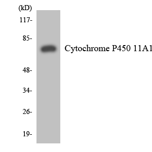
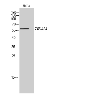
Cn|En

现货抗体产品库

Cyclin D1 Rabbit Polyclonal Antibody

产品详情 相关文献 产品问答 相关产品

产品基本信息

产品货号
BD-PT1173
产品名称
Cyclin D1 Rabbit Polyclonal Antibody
别名
CCND1; BCL1; PRAD1; G1/S-specific cyclin-D1; B-cell lymphoma 1 protein; BCL-1; BCL-1 oncogene; PRAD1 oncogene
类别
常规抗体
基因名称
CCND1
蛋白名称
G1/S-specific cyclin-D1
推荐应用
WB
反应种属
Human,Mouse,Rat,Pig
浓度
1 mg/ml
存储缓冲液
Liquid in PBS containing 50% glycerol, 0.5% BSA and 0.02% New type preservative N.
Human Gene ID
595
Human Gene Link
http://www.ncbi.nlm.nih.gov/sites/entrez?db=gene&term=595
Human Swissprot No.
P24385
Human Swissprot Link
http://www.uniprot.org/uniprotkb/P24385/entry
Mouse Gene ID
12443
Mouse Gene Link
http://www.ncbi.nlm.nih.gov/sites/entrez?db=gene&term=12443
Mouse Swissprot No.
P25322
Mouse Swissprot Link
http://www.uniprot.org/uniprot/P25322
Rat Gene ID
58919
Rat Gene Link
http://www.ncbi.nlm.nih.gov/sites/entrez?db=gene&term=58919
Rat Swissprot No.
P39948
Rat Swissprot Link
http://www.uniprot.org/uniprot/P39948
免疫原
The antiserum was produced against synthesized peptide derived from human Cyclin D1. AA range:246-295
特异性
Cyclin D1 Polyclonal Antibody detects endogenous levels of Cyclin D1 protein.
稀释度
IF 1:50-200 WB 1:500 - 1:2000. IHC 1:100 - 1:300. ELISA: 1:40000. Not yet tested in other applications.
预测分子量
33kD
运输及保存条件
-20°C/1 year
宿主
Polyclonal, Rabbit,IgG
背景介绍
The protein encoded by this gene belongs to the highly conserved cyclin family, whose members are characterized by a dramatic periodicity in protein abundance throughout the cell cycle. Cyclins function as regulators of CDK kinases. Different cyclins exhibit distinct expression and degradation patterns which contribute to the temporal coordination of each mitotic event. This cyclin forms a complex with and functions as a regulatory subunit of CDK4 or CDK6, whose activity is required for cell cycle G1/S transition. This protein has been shown to interact with tumor suppressor protein Rb and the expression of this gene is regulated positively by Rb. Mutations, amplification and overexpression of this gene, which alters cell cycle progression, are observed frequently in a variety of tumors and may contribute to tumorigenesis. [provided by RefSeq, Jul 2008],
组织表达
Brain,Placenta,Tongue,
细胞定位
Nucleus . Cytoplasm . Nucleus membrane . Cyclin D-CDK4 complexes accumulate at the nuclear membrane and are then translocated to the nucleus through interaction with KIP/CIP family members. .
信号通路
Cell_Cycle_G1S;Cell_Cycle_G2M_DNA;p53;WNT;WNT-T CELLFocal adhesion;Jak_STAT;Pathways in cancer;Colorectal cancer;Pancreatic cancer;Endometrial cancer;Glioma;Prostate cancer;Thyroid cancer;Melanoma;Bladder cancer;Chronic myeloid leukemia;Acute myeloid leuk
功能
disease:A chromosomal aberration involving CCND1 may be a cause of B-lymphocytic malignancy, particularly mantle-cell lymphoma (MCL). Translocation t(11;14)(q13;q32) with immunoglobulin gene regions. Activation of CCND1 may be oncogenic by directly altering progression through the cell cycle.,disease:A chromosomal aberration involving CCND1 may be a cause of multiple myeloma [MIM:254500]. Translocation t(11;14)(q13;q32) with the IgH locus.,disease:A chromosomal aberration involving CCND1 may be a cause of parathyroid adenomas [MIM:168461]. Translocation t(11;11)(q13;p15) with the parathyroid hormone (PTH) enhancer.,function:Essential for the control of the cell cycle at the G1/S (start) transition.,online information:The Singapore human mutation and polymorphism database,PTM:Following DNA damage it is ubiquitinated by some SCF (SKP1-cullin-F-box) protein ligase complex containing FBXO31. Ubiquitination leads to its degradation and G1 arrest.,PTM:Phosphorylation at Thr-286 by MAP kinases is required for ubiquitination and degradation following DNA damage. It probably plays an essential role for recognition by the FBXO31 component of SCF (SKP1-cullin-F-box) protein ligase complex.,similarity:Belongs to the cyclin family.,similarity:Belongs to the cyclin family. Cyclin D subfamily.,subunit:Interacts with the CDK4 and CDK6 protein kinases to form a serine/threonine kinase holoenzyme complex. The cyclin subunit imparts substrate specificity to the complex.,
期货
现货
纯化
The antibody was affinity-purified from rabbit antiserum by affinity-chromatography using epitope-specific immunogen.

Immunohistochemistry analysis of paraffin-embedded human brain tissue, using Cyclin D1 Antibody. The picture on the right is blocked with the synthesized peptide.

Western blot analysis of lysates from Jurkat cells, treated with EGF 200ng/ml 30‘, using Cyclin D1 Antibody. The lane on the right is blocked with the synthesized peptide.

Immunofluorescence analysis of mouse-spleen tissue. 1,Cyclin D1 Polyclonal Antibody(red) was diluted at 1:200(4°C,overnight). 2, Cy3 labled Secondary antibody was diluted at 1:300(room temperature, 50min).3, Picture B: DAPI(blue) 10min. Picture A:Target. Picture B: DAPI. Picture C: merge of A+B

Immunofluorescence analysis of rat-spleen tissue. 1,Cyclin D1 Polyclonal Antibody(red) was diluted at 1:200(4°C,overnight). 2, Cy3 labled Secondary antibody was diluted at 1:300(room temperature, 50min).3, Picture B: DAPI(blue) 10min. Picture A:Target. Picture B: DAPI. Picture C: merge of A+B

Immunohistochemical analysis of paraffin-embedded Rat-spleen tissue. 1,Cyclin D1 Polyclonal Antibody was diluted at 1:200(4°C,overnight). 2, Sodium citrate pH 6.0 was used for antibody retrieval(>98°C,20min). 3,Secondary antibody was diluted at 1:200(room tempeRature, 30min). Negative control was used by secondary antibody only.

Immunohistochemical analysis of paraffin-embedded Mouse-lung tissue. 1,Cyclin D1 Polyclonal Antibody was diluted at 1:200(4°C,overnight). 2, Sodium citrate pH 6.0 was used for antibody retrieval(>98°C,20min). 3,Secondary antibody was diluted at 1:200(room tempeRature, 30min). Negative control was used by secondary antibody only.

Western Blot analysis of various cells using Cyclin D1 Polyclonal Antibody diluted at 1:1000

Western Blot analysis of MCF-7 cells using Cyclin D1 Polyclonal Antibody diluted at 1:1000

Immunohistochemical analysis of paraffin-embedded Human brain. Antibody was diluted at 1:100(4°,overnight). High-pressure and temperature Tris-EDTA,pH8.0 was used for antigen retrieval. Negetive contrl (right) obtaned from antibody was pre-absorbed by immunogen peptide.

Immunohistochemical analysis of paraffin-embedded Human skin. 1, Antibody was diluted at 1:100(4°,overnight). 2, High-pressure and temperature EDTA, pH8.0 was used for antigen retrieval. 3,Secondary antibody was diluted at 1:200(room temperature, 30min).

Immunohistochemical analysis of paraffin-embedded Human skin. 1, Antibody was diluted at 1:100(4°,overnight). 2, High-pressure and temperature EDTA, pH8.0 was used for antigen retrieval. 3,Secondary antibody was diluted at 1:200(room temperature, 30min).

Immunohistochemical analysis of paraffin-embedded Human skin. 1, Antibody was diluted at 1:100(4°,overnight). 2, High-pressure and temperature EDTA, pH8.0 was used for antigen retrieval. 3,Secondary antibody was diluted at 1:200(room temperature, 30min).

相关文献

产品问答

相关产品

免责声明| 法律支持| 联系方式

市场:027-65023363   行政/人事:027-62439686   邮箱:marketing@brainvta.com  

销售总监:张经理  18995532642  华东区:陈经理 18013970337   华南区:王经理 13100653525   华中/西区:杨经理 18186518905   华北区:张经理 18893721749

地址:中国武汉东湖高新区光谷七路128号中科开物产业园1号楼

Copyright © 武汉枢密脑科学技术有限公司. All RIGHTS RESERVED.
鄂ICP备2021009124号 DIGITAL BY VTHINK