CTPS Rabbit Polyclonal Antibody
产品基本信息
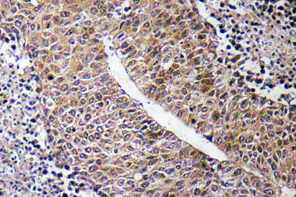
Immunohistochemistry analysis of CTPS antibody in paraffin-embedded human liver carcinoma tissue.
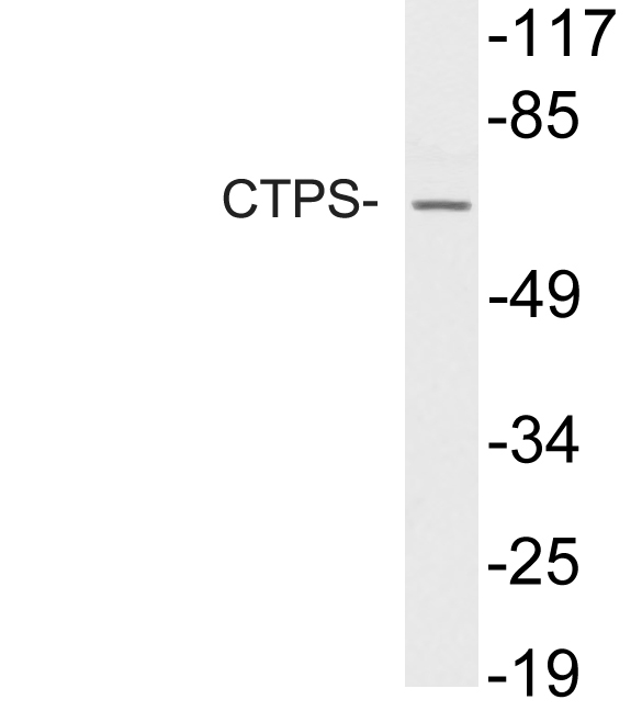
Western blot analysis of lysate from HUVEC cells, using CTPS antibody.
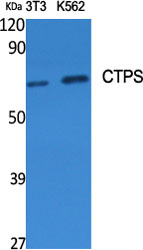
Western Blot analysis of various cells using CTPS Polyclonal Antibody diluted at 1:500
.jpg)
Western Blot analysis of HuvEc cells using CTPS Polyclonal Antibody diluted at 1:500
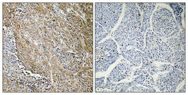
Immunohistochemical analysis of paraffin-embedded Human lung cancer. Antibody was diluted at 1:100(4°,overnight). High-pressure and temperature Tris-EDTA,pH8.0 was used for antigen retrieval. Negetive contrl (right) obtaned from antibody was pre-absorbed by immunogen peptide.
相关文献
产品问答
相关产品

市场:027-65023363 行政/人事:027-62439686 邮箱:marketing@brainvta.com 客服:18140661572(活动咨询、售后反馈等)
销售总监:张经理 18995532642 华东区:陈经理 18013970337 华南区:王经理 13100653525 华中/西区:杨经理 18186518905 华北区:张经理 18893721749
地址:中国武汉东湖高新区光谷七路128号中科开物产业园1号楼
Copyright © 武汉枢密脑科学技术有限公司. All RIGHTS RESERVED.
鄂ICP备2021009124号 DIGITAL BY VTHINK
