产品名称
Cofilin Rabbit Polyclonal Antibody
别名
CFL1; CFL; Cofilin-1; 18 kDa phosphoprotein; p18; Cofilin; non-muscle isoform
存储缓冲液
Liquid in PBS containing 50% glycerol, 0.5% BSA and 0.02% New type preservative N.
Human Gene Link
http://www.ncbi.nlm.nih.gov/sites/entrez?db=gene&term=1072
Human Swissprot No.
P23528
Human Swissprot Link
http://www.uniprot.org/uniprotkb/P23528/entry
Mouse Gene Link
http://www.ncbi.nlm.nih.gov/sites/entrez?db=gene&term=12631
Mouse Swissprot No.
P18760
Mouse Swissprot Link
http://www.uniprot.org/uniprot/P18760
Rat Gene Link
http://www.ncbi.nlm.nih.gov/sites/entrez?db=gene&term=29271
Rat Swissprot Link
http://www.uniprot.org/uniprot/P45592
免疫原
The antiserum was produced against synthesized peptide derived from human Cofilin. AA range:1-50
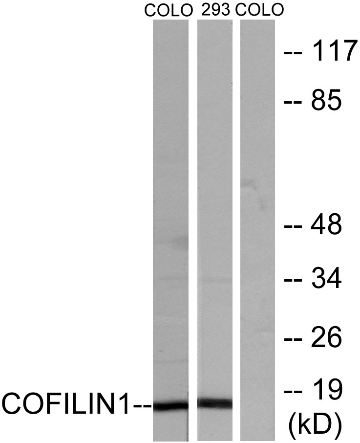
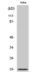
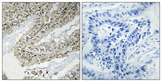
特异性
Cofilin Polyclonal Antibody detects endogenous levels of Cofilin protein.
宿主
Polyclonal, Rabbit,IgG
背景介绍
cofilin 1(CFL1) Homo sapiens The protein encoded by this gene can polymerize and depolymerize F-actin and G-actin in a pH-dependent manner. Increased phosphorylation of this protein by LIM kinase aids in Rho-induced reorganization of the actin cytoskeleton. Cofilin is a widely distributed intracellular actin-modulating protein that binds and depolymerizes filamentous F-actin and inhibits the polymerization of monomeric G-actin in a pH-dependent manner. It is involved in the translocation of actin-cofilin complex from cytoplasm to nucleus.[supplied by OMIM, Apr 2004],
组织表达
Widely distributed in various tissues.
细胞定位
Nucleus matrix . Cytoplasm, cytoskeleton . Cell projection, ruffle membrane ; Peripheral membrane protein ; Cytoplasmic side . Cell projection, lamellipodium membrane ; Peripheral membrane protein ; Cytoplasmic side . Cell projection, lamellipodium . Cell projection, growth cone . Cell projection, axon . Colocalizes with the actin cytoskeleton in membrane ruffles and lamellipodia. Detected at the cleavage furrow and contractile ring during cytokinesis. Almost completely in nucleus in cells exposed to heat shock or 10% dimethyl sulfoxide.
信号通路
Axon guidance;Fc gamma R-mediated phagocytosis;Regulates Actin and Cytoskeleton;
功能
function:Controls reversibly actin polymerization and depolymerization in a pH-sensitive manner. It has the ability to bind G- and F-actin in a 1:1 ratio of cofilin to actin. It is the major component of intranuclear and cytoplasmic actin rods.,online information:Cofilin entry,PTM:Phosphorylated on Ser-3 in resting cells.,similarity:Belongs to the actin-binding proteins ADF family.,similarity:Contains 1 ADF-H domain.,subcellular location:Almost completely in nucleus in cells exposed to heat shock or 10% dimethyl sulfoxide.,tissue specificity:Widely distributed in various tissues.,
纯化
The antibody was affinity-purified from rabbit antiserum by affinity-chromatography using epitope-specific immunogen.