产品名称
Centrobin Rabbit Polyclonal Antibody
别名
CNTROB; LIP8; PP1221; Centrobin; Centrosomal BRCA2-interacting protein; LYST-interacting protein 8
存储缓冲液
Liquid in PBS containing 50% glycerol, 0.5% BSA and 0.02% New type preservative N.
Human Gene Link
http://www.ncbi.nlm.nih.gov/sites/entrez?db=gene&term=116840
Human Swissprot No.
Q8N137
Human Swissprot Link
http://www.uniprot.org/uniprotkb/Q8N137/entry
Mouse Swissprot No.
Q8CB62
Mouse Swissprot Link
http://www.uniprot.org/uniprot/Q8CB62
免疫原
The antiserum was produced against synthesized peptide derived from human CNTROB. AA range:591-640
特异性
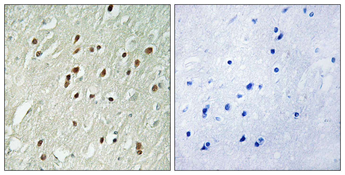
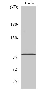
Centrobin Polyclonal Antibody detects endogenous levels of Centrobin protein.
宿主
Polyclonal, Rabbit,IgG
背景介绍
This gene encodes a centrosomal protein that interacts with BRCA2, and is required for centriole duplication and cytokinesis. Alternatively spliced transcript variants encoding different isoforms have been described for this gene. [provided by RefSeq, Aug 2011],
组织表达
Widely expressed (at protein level). Highly expressed in testis. Also expressed in spleen, thymus, prostate, small intestine, colon and peripheral blood leukocytes.
细胞定位
Cytoplasm, cytoskeleton, microtubule organizing center, centrosome, centriole . Centriole-associated, asymmetrically localizes to the daughter centriole.
功能
developmental stage:Preferentially incorporated into the newly assembled daughter centriole during centriole assembly at the late G1 or early S phase. Remains in the daughter centrioles throughout the cell cycle. At the next cycle of centriole duplication, its amount on the original daughter centriole eventually decreases.,function:Required for centriole duplication. Inhibition of centriole duplication leading to defects in cytokinesis.,PTM:Phosphorylated upon DNA damage, probably by ATM or ATR.,sequence caution:Wrong choice of frame.,subcellular location:Centriole-associated, asymmetrically localizes to the daughter centriole.,subunit:Interacts with LYST.,tissue specificity:Widely expressed (at protein level). Highly expressed in testis. Also expressed in spleen, thymus, prostate, small intestine, colon and peripheral blood leukocytes.,
纯化
The antibody was affinity-purified from rabbit antiserum by affinity-chromatography using epitope-specific immunogen.