产品名称
CaMKIIα/δ Rabbit Polyclonal Antibody
别名
CAMK2A; CAMKA; KIAA0968; Calcium/calmodulin-dependent protein kinase type II subunit alpha; CaM kinase II subunit alpha; CaMK-II subunit alpha; CAMK2D; CAMKD; Calcium/calmodulin-dependent protein kinase type II subunit delta; CaM kinase II
蛋白名称
Calcium/calmodulin-dependent protein kinase type II subunit alpha/delta
存储缓冲液
Liquid in PBS containing 50% glycerol, 0.5% BSA and 0.02% New type preservative N.
Human Gene Link
http://www.ncbi.nlm.nih.gov/sites/entrez?db=gene&term=815
Human Swissprot No.
Q9UQM7/Q13557
Human Swissprot Link
http://www.uniprot.org/uniprotkb/Q9UQM7/entry
Mouse Gene ID
12322/108058
Mouse Gene Link
http://www.ncbi.nlm.nih.gov/sites/entrez?db=gene&term=12322
Rat Gene Link
http://www.ncbi.nlm.nih.gov/sites/entrez?db=gene&term=25400
Rat Swissprot No.
P11275/P15791
Rat Swissprot Link
http://www.uniprot.org/uniprot/P11275
免疫原
The antiserum was produced against synthesized peptide derived from human CaMK2 alpha/delta. AA range:256-305
特异性
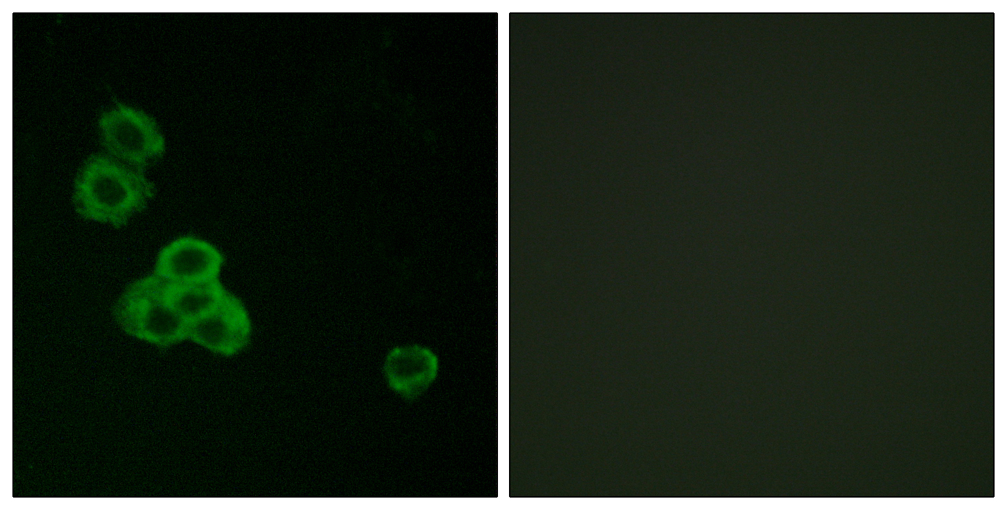
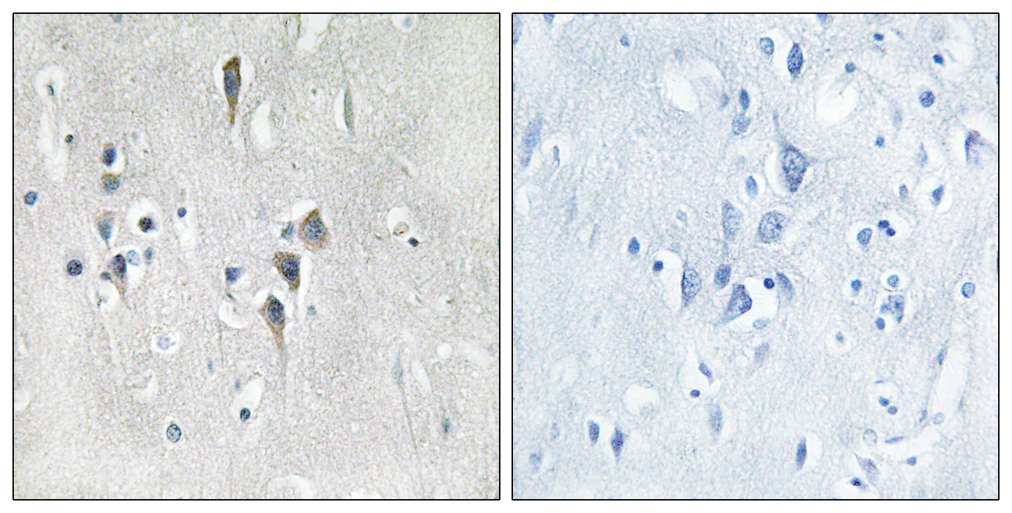
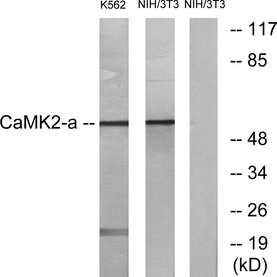
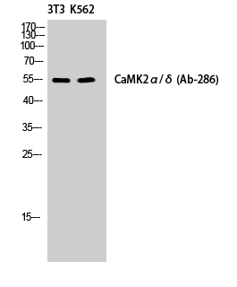
CaMKIIα/δ Polyclonal Antibody detects endogenous levels of CaMKIIα/δ protein.
宿主
Polyclonal, Rabbit,IgG
背景介绍
The product of this gene belongs to the serine/threonine protein kinases family, and to the Ca(2+)/calmodulin-dependent protein kinases subfamily. Calcium signaling is crucial for several aspects of plasticity at glutamatergic synapses. This calcium calmodulin-dependent protein kinase is composed of four different chains: alpha, beta, gamma, and delta. The alpha chain encoded by this gene is required for hippocampal long-term potentiation (LTP) and spatial learning. In addition to its calcium-calmodulin (CaM)-dependent activity, this protein can undergo autophosphorylation, resulting in CaM-independent activity. Two transcript variants encoding distinct isoforms have been identified for this gene. [provided by RefSeq, Nov 2008],
细胞定位
Cell junction, synapse . Cell junction, synapse, postsynaptic density . Cell projection, dendritic spine . Cell projection, dendrite . Postsynaptic lipid rafts. .
信号通路
ErbB_HER;Calcium;Oocyte meiosis;WNT;WNT-T CELLLong-term potentiation;Neurotrophin;Olfactory transduction;GnRH;Melanogenesis;Glioma;
功能
catalytic activity:ATP + a protein = ADP + a phosphoprotein.,enzyme regulation:Autophosphorylation of Thr-286 allows the kinase to switch from a calmodulin-dependent to a calmodulin-independent state.,function:CaM-kinase II (CAMK2) is a prominent kinase in the central nervous system that may function in long-term potentiation and neurotransmitter release. Member of the NMDAR signaling complex in excitatory synapses it may regulate NMDAR-dependent potentiation of the AMPAR and synaptic plasticity.,similarity:Belongs to the protein kinase superfamily.,similarity:Belongs to the protein kinase superfamily. CAMK Ser/Thr protein kinase family. CaMK subfamily.,similarity:Contains 1 protein kinase domain.,subcellular location:Postsynaptic lipid rafts.,subunit:CAMK2 is composed of four different chains: alpha, beta, gamma, and delta. The different isoforms assemble into homo- or heteromultimeric holoenzymes composed of 8 to 12 subunits. Interacts with BAALC, MPDZ, SYN1, CAMK2N2 and SYNGAP1.,
纯化
The antibody was affinity-purified from rabbit antiserum by affinity-chromatography using epitope-specific immunogen.