产品名称
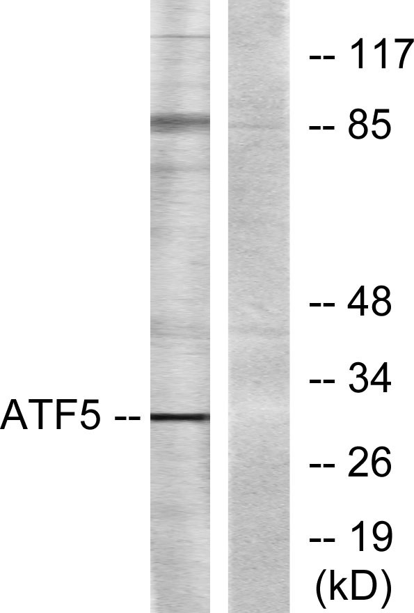
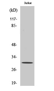
ATF-5 Rabbit Polyclonal Antibody
别名
ATF5; ATFX; Cyclic AMP-dependent transcription factor ATF-5; cAMP-dependent transcription factor ATF-5; Activating transcription factor 5; Transcription factor ATFx
蛋白名称
Cyclic AMP-dependent transcription factor ATF-5
存储缓冲液
Liquid in PBS containing 50% glycerol, 0.5% BSA and 0.02% New type preservative N.
Human Gene Link
http://www.ncbi.nlm.nih.gov/sites/entrez?db=gene&term=22809
Human Swissprot No.
Q9Y2D1
Human Swissprot Link
http://www.uniprot.org/uniprotkb/Q9Y2D1/entry
Mouse Gene Link
http://www.ncbi.nlm.nih.gov/sites/entrez?db=gene&term=107503
Mouse Swissprot No.
O70191
Mouse Swissprot Link
http://www.uniprot.org/uniprot/O70191
Rat Gene Link
http://www.ncbi.nlm.nih.gov/sites/entrez?db=gene&term=282840
Rat Swissprot Link
http://www.uniprot.org/uniprot/Q6P788
免疫原
The antiserum was produced against synthesized peptide derived from human ATF5. AA range:221-270
特异性
ATF-5 Polyclonal Antibody detects endogenous levels of ATF-5 protein.
宿主
Polyclonal, Rabbit,IgG
背景介绍
function:Transcriptional activator which binds the cAMP response element (CRE) (consensus: 5'-GTGACGT[AC][AG]-3'), a sequence present in many viral and cellular promoters and blocks the differentiation of neuroprogenitor cells into neurons. Its transcriptional activity is enhanced by CCND3 and slightly inhibited by CDK4.,similarity:Belongs to the bZIP family.,similarity:Contains 1 bZIP domain.,subunit:Binds DNA as a dimer. Interacts with PTP4A1/PRL-1 (By similarity). Interacts with CCND3, but not with CCND1 or CCND2.,
组织表达
Widely expressed with higher expression levels in liver.
细胞定位
Cytoplasm . Nucleus . Cytoplasm, cytoskeleton, microtubule organizing center, centrosome . Actively transported to the centrosome and accumulated in the pericentriolar material (PCM) during G1 to M phase via a microtubule-dependent mechanism. During late telophase and cytokinesis, translocates from the centrosome to the midbody. .
功能
function:Transcriptional activator which binds the cAMP response element (CRE) (consensus: 5'-GTGACGT[AC][AG]-3'), a sequence present in many viral and cellular promoters and blocks the differentiation of neuroprogenitor cells into neurons. Its transcriptional activity is enhanced by CCND3 and slightly inhibited by CDK4.,similarity:Belongs to the bZIP family.,similarity:Contains 1 bZIP domain.,subunit:Binds DNA as a dimer. Interacts with PTP4A1/PRL-1 (By similarity). Interacts with CCND3, but not with CCND1 or CCND2.,
纯化
The antibody was affinity-purified from rabbit antiserum by affinity-chromatography using epitope-specific immunogen.