产品名称
ATF-1 Rabbit Polyclonal Antibody
别名
ATF1; Cyclic AMP-dependent transcription factor ATF-1; cAMP-dependent transcription factor ATF-1; Activating transcription factor 1; Protein TREB36
蛋白名称
Cyclic AMP-dependent transcription factor ATF-1
存储缓冲液
Liquid in PBS containing 50% glycerol, 0.5% BSA and 0.02% New type preservative N.
Human Gene Link
http://www.ncbi.nlm.nih.gov/sites/entrez?db=gene&term=466
Human Swissprot No.
P18846
Human Swissprot Link
http://www.uniprot.org/uniprotkb/P18846/entry
Mouse Gene Link
http://www.ncbi.nlm.nih.gov/sites/entrez?db=gene&term=11908
Mouse Swissprot No.
P81269
Mouse Swissprot Link
http://www.uniprot.org/uniprot/P81269
免疫原
The antiserum was produced against synthesized peptide derived from human ATF1. AA range:31-80
特异性
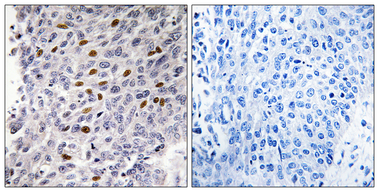
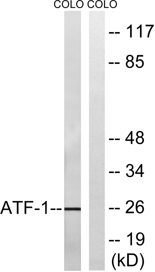
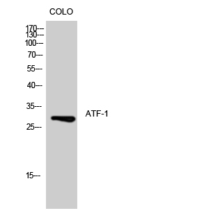
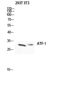
ATF-1 Polyclonal Antibody detects endogenous levels of ATF-1 protein.
宿主
Polyclonal, Rabbit,IgG
背景介绍
activating transcription factor 1(ATF1) Homo sapiens This gene encodes an activating transcription factor, which belongs to the ATF subfamily and bZIP (basic-region leucine zipper) family. It influences cellular physiologic processes by regulating the expression of downstream target genes, which are related to growth, survival, and other cellular activities. This protein is phosphorylated at serine 63 in its kinase-inducible domain by serine/threonine kinases, cAMP-dependent protein kinase A, calmodulin-dependent protein kinase I/II, mitogen- and stress-activated protein kinase and cyclin-dependent kinase 3 (cdk-3). Its phosphorylation enhances its transactivation and transcriptional activities, and enhances cell transformation. Fusion of this gene and FUS on chromosome 16 or EWSR1 on chromosome 22 induced by translocation generates chimeric proteins in angiomatoid fibrous histiocytoma and clear cell sarcoma. This gene has a pseudogene on chro
功能
disease:A chromosomal aberration involving ATF1 is associated with angiomatoid fibrous histiocytoma (AFH) [MIM:612160]. Translocation t(12;16)(q13;p11.2) with FUS generates a chimeric ATF1/FUS protein.,disease:A chromosomal aberration involving ATF1 is associated with angiomatoid fibrous histiocytoma (AFH) [MIM:612160]. Translocation t(12;22)(q13;q12) with EWSR1 generates a chimeric ATF1/EWSR1 protein.,function:This protein binds the cAMP response element (CRE) (consensus: 5'-GTGACGT[AC][AG]-3'), a sequence present in many viral and cellular promoters. Binds to the Tax-responsive element (TRE) of HTLV-I. Mediates PKA-induced stimulation of CRE-reporter genes.,similarity:Belongs to the bZIP family. ATF subfamily.,similarity:Contains 1 bZIP domain.,similarity:Contains 1 KID (kinase-inducible) domain.,subunit:Binds DNA as a dimer.,
纯化
The antibody was affinity-purified from rabbit antiserum by affinity-chromatography using epitope-specific immunogen.