产品名称
APLP-2 Rabbit Polyclonal Antibody
别名
APLP2; APPL2; Amyloid-like protein 2; APLP-2; APPH; Amyloid protein homolog; CDEI box-binding protein; CDEBP
蛋白名称
Amyloid-like protein 2
存储缓冲液
Liquid in PBS containing 50% glycerol, 0.5% BSA and 0.02% New type preservative N.
Human Gene Link
http://www.ncbi.nlm.nih.gov/sites/entrez?db=gene&term=334
Human Swissprot No.
Q06481
Human Swissprot Link
http://www.uniprot.org/uniprotkb/Q06481/entry
Mouse Gene Link
http://www.ncbi.nlm.nih.gov/sites/entrez?db=gene&term=11804
Mouse Swissprot No.
Q06335
Mouse Swissprot Link
http://www.uniprot.org/uniprot/Q06335
Rat Swissprot Link
http://www.uniprot.org/uniprot/P15943
免疫原
The antiserum was produced against synthesized peptide derived from human APLP2. AA range:241-290
特异性
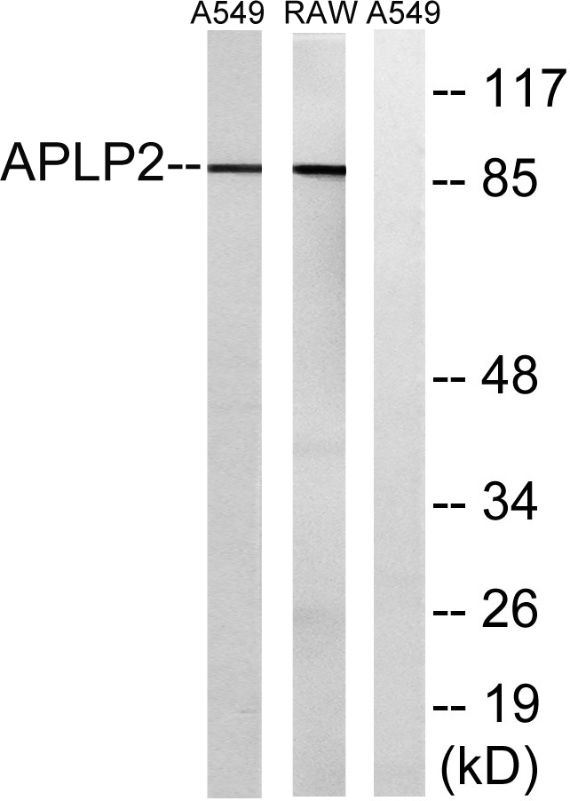
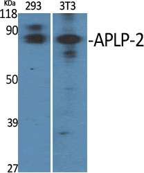
APLP-2 Polyclonal Antibody detects endogenous levels of APLP-2 protein.
宿主
Polyclonal, Rabbit,IgG
背景介绍
This gene encodes amyloid precursor- like protein 2 (APLP2), which is a member of the APP (amyloid precursor protein) family including APP, APLP1 and APLP2. This protein is ubiquitously expressed. It contains heparin-, copper- and zinc- binding domains at the N-terminus, BPTI/Kunitz inhibitor and E2 domains in the middle region, and transmembrane and intracellular domains at the C-terminus. This protein interacts with major histocompatibility complex (MHC) class I molecules. The synergy of this protein and the APP is required to mediate neuromuscular transmission, spatial learning and synaptic plasticity. This protein has been implicated in the pathogenesis of Alzheimer's disease. Multiple alternatively spliced transcript variants encoding different isoforms have been identified. [provided by RefSeq, Aug 2011],
组织表达
Expressed in placenta, brain, heart, lung, liver, kidney and endothelial tissues.
细胞定位
Cell membrane ; Single-pass type I membrane protein . Nucleus .
功能
alternative products:Additional isoforms seem to exist,function:May play a role in the regulation of hemostasis. The soluble form may have inhibitory properties towards coagulation factors. May interact with cellular G-protein signaling pathways. May bind to the DNA 5'-GTCACATG-3'(CDEI box). Inhibits trypsin, chymotrypsin, plasmin, factor XIA and plasma and glandular kallikrein.,PTM:The BPTI/Kunitz inhibitor domain is O-glycosylated.,similarity:Belongs to the APP family.,similarity:Contains 1 BPTI/Kunitz inhibitor domain.,subunit:Interacts with CPEB1.,tissue specificity:In placenta, brain, heart, lung, liver, kidney and endothelial tissues.,
纯化
The antibody was affinity-purified from rabbit antiserum by affinity-chromatography using epitope-specific immunogen.
.jpg)

