产品名称
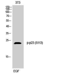
p23 (phospho Ser113) Rabbit Polyclonal Antibody
别名
PTGES3; P23; TEBP; Prostaglandin E synthase 3; Cytosolic prostaglandin E2 synthase; cPGES; Hsp90 co-chaperone; Progesterone receptor complex p23; Telomerase-binding protein p23
蛋白名称
Prostaglandin E synthase 3
存储缓冲液
Liquid in PBS containing 50% glycerol, 0.5% BSA and 0.02% New type preservative N.
Human Gene Link
http://www.ncbi.nlm.nih.gov/sites/entrez?db=gene&term=10728
Human Swissprot No.
Q15185
Human Swissprot Link
http://www.uniprot.org/uniprotkb/Q15185/entry
Mouse Gene Link
http://www.ncbi.nlm.nih.gov/sites/entrez?db=gene&term=100043508
Mouse Swissprot No.
Q9R0Q7
Mouse Swissprot Link
http://www.uniprot.org/uniprot/Q9R0Q7
Rat Gene Link
http://www.ncbi.nlm.nih.gov/sites/entrez?db=gene&term=362809
Rat Swissprot Link
http://www.uniprot.org/uniprot/P83868
免疫原
The antiserum was produced against synthesized peptide derived from human TEBP around the phosphorylation site of Ser113. AA range:79-128
特异性
Phospho-p23 (S113) Polyclonal Antibody detects endogenous levels of p23 protein only when phosphorylated at S113.
宿主
Polyclonal, Rabbit,IgG
背景介绍
This gene encodes an enzyme that converts prostaglandin endoperoxide H2 (PGH2) to prostaglandin E2 (PGE2). This protein functions as a co-chaperone with heat shock protein 90 (HSP90), localizing to response elements in DNA and disrupting transcriptional activation complexes. Alternative splicing results in multiple transcript variants. There are multiple pseudogenes of this gene on several different chromosomes. [provided by RefSeq, Feb 2016],
组织表达
Embryonic kidney,Epithelium,Liver,Lung,Pituitary,Platelet,T-cell,Testis,Uri
功能
catalytic activity:(5Z,13E)-(15S)-9-alpha,11-alpha-epidioxy-15-hydroxyprosta-5,13-dienoate = (5Z,13E)-(15S)-11-alpha,15-dihydroxy-9-oxoprosta-5,13-dienoate.,function:Molecular chaperone that localizes to genomic response elements in a hormone-dependent manner and disrupts receptor-mediated transcriptional activation, by promoting disassembly of transcriptional regulatory complexes.,pathway:Lipid metabolism; prostaglandin biosynthesis.,similarity:Belongs to the p23/wos2 family.,similarity:Contains 1 CS domain.,subunit:Binds to telomerase and to the progesterone receptor.,
纯化
The antibody was affinity-purified from rabbit antiserum by affinity-chromatography using epitope-specific immunogen.