P40 (1X6) Rabbit Monoclonal antibody
产品基本信息
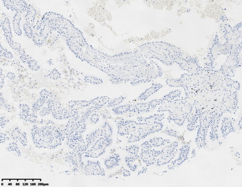
Immunohistochemical staining of paraffin-embedded human lung adenocarcinoma tissue using anti-P40 (ΔNp63) rabbit monoclonal antibody (RM1400). Heat-induced epitope retrieval by 1mM EDTA in 10mM Tris buffer (pH9.0) at 120°C for 3 min. (1:35000)
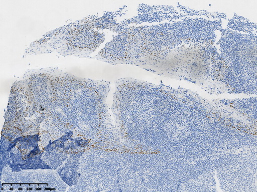
Immunohistochemical staining of paraffin-embedded Human tonsil within the normal limits using anti-P40 (ΔNp63) rabbit monoclonal antibody (RM1400). Heat-induced epitope retrieval by 1mM EDTA in 10mM Tris buffer (pH9.0) at 120°C for 3 min. (1:35000)
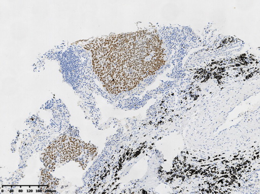
Immunohistochemical staining of paraffin-embedded human lung?squamous?cell?carcinoma tissue using anti-P40 (ΔNp63) rabbit monoclonal antibody (RM1400). Heat-induced epitope retrieval by 1mM EDTA in 10mM Tris buffer (pH9.0) at 120°C for 3 min. (1:35000)
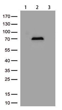
Western blot analysis of lysates from HEK293T cells transfected with P40 (ΔNp63) (Lane 2), TP63 overexpression plasmid (Lane 3) or empty vector plasmid (Lane 1) using anti-P40 (ΔNp63) antibody (Cat# RM1400). (1:1000)
相关文献
产品问答
相关产品

市场:027-65023363 行政/人事:027-62439686 邮箱:marketing@brainvta.com 客服:18140661572(活动咨询、售后反馈等)
销售总监:张经理 18995532642 华东区:陈经理 18013970337 华南区:王经理 13100653525 华中/西区:杨经理 18186518905 华北区:张经理 18893721749
地址:中国武汉东湖高新区光谷七路128号中科开物产业园1号楼
Copyright © 武汉枢密脑科学技术有限公司. All RIGHTS RESERVED.
鄂ICP备2021009124号 DIGITAL BY VTHINK
