Cn|En

现货抗体产品库

SDCBP (17Z12) Mouse Monoclonal antibody

产品详情 相关文献 产品问答 相关产品

产品基本信息

产品货号
BD-PE2721
产品名称
SDCBP (17Z12) Mouse Monoclonal antibody
类别
常规抗体
基因名称
SDCBP
推荐应用
WB
反应种属
Human,Mouse,Rat
存储缓冲液
PBS (pH 7.3) containing 1% BSA, 50% glycerol and 0.02% New type preservative N.
免疫原
Full length human recombinant protein of human SDCBP(NP_005616) produced in HEK293T cell.
特异性
Validated through 10k CHIP testing,using a High-Density Protein Microarray that was spotted with ~17,000 various over-expressed proteins to test for antibody specificity.
稀释度
WB 1:500, IHC 1:100
预测分子量
32.3kDa
运输及保存条件
-20°C/1 year
宿主
Mouse
同种型
IgG2b
背景介绍
The protein encoded by this gene was initially identified as a molecule linking syndecan-mediated signaling to the cytoskeleton. The syntenin protein contains tandemly repeated PDZ domains that bind the cytoplasmic, C-terminal domains of a variety of transmembrane proteins. This protein may also affect cytoskeletal-membrane organization, cell adhesion, protein trafficking, and the activation of transcription factors. The protein is primarily localized to membrane-associated adherens junctions and focal adhesions but is also found at the endoplasmic reticulum and nucleus. Alternative splicing results in multiple transcript variants encoding different isoforms. [provided by RefSeq, Jul 2008]
期货
现货
纯化
Purified from mouse ascites fluids or tissue culture supernatant by affinity chromatography (protein A/G)

OriGene overexpression protein microarray chip was immunostained with UltraMAB anti-SDCBP mouse monoclonal antibody (BD-PE2721). The positive reactive proteins are highlighted with two red arrows in the enlarged subarray. All the positive controls spotted in this subarray are also labeled for clarification.

Immunohistochemical staining of paraffin-embedded composit of 2 human melanoma cases using anti-SDCBP 9 mouse monoclonal antibody (BD-PE2721) at 1:100 with Polink2 Broad HRP DAB detection kit; heat-induced epitope retrieval with GBI Citrate pH6.0 HIER buffer using pressure chamber for 3 minutes at 110C. Membrane, nuclear, and cytoplasmic staining is seen in tumor cells with various intensity.

Immunohistochemical staining of paraffin-embedded tonsil using anti-SDCBP 9 mouse monoclonal antibody (BD-PE2721) at 1:100 with Polink2 Broad HRP DAB detection kit; heat-induced epitope retrieval with GBI Citrate pH6.0 HIER buffer using pressure chamber for 3 minutes at 110C. Membrane and cytoplasmic staining is seen in germinal and non germinal center of the the tonsil.

Immunohistochemical staining of paraffin-embedded mouse liver tissue using anti-SDCBP (SYNTINEN) 9 mouse monoclonal antibody. HIER TEE buffer pH9 at 110C for 10 min, BD-PE2721 (1:100). Detection was done with Klear Mouse (C/N ) DAB Kit.

Immunohistochemical staining of paraffin-embedded human brain using anti-SDCBP 9 mouse monoclonal antibody (BD-PE2721) at 1:100 with Polink2 Broad HRP DAB detection kit; heat-induced epitope retrieval with GBI Citrate pH6.0 HIER buffer using pressure chamber for 3 minutes at 110C. Weak membrane and cytoplasmic staining.

Immunohistochemical staining of paraffin-embedded composit of 3 human endometrial cancer cases using anti-SDCBP 9 mouse monoclonal antibody (BD-PE2721) at 1:100 with Polink2 Broad HRP DAB detection kit; heat-induced epitope retrieval with GBI Citrate pH6.0 HIER buffer using pressure chamber for 3 minutes at 110C. Membrane and cytoplasmic staining is seen in tumor cells with various intensity.

Immunohistochemical staining of paraffin-embedded composit of 3 human liver cancer cases using anti-SDCBP 9 mouse monoclonal antibody (BD-PE2721) at 1:100 with Polink2 Broad HRP DAB detection kit; heat-induced epitope retrieval with GBI Citrate pH6.0 HIER buffer using pressure chamber for 3 minutes at 110C. Membrane and cytoplasmic staining is seen in tumor cells with various intensity.

Immunohistochemical staining of paraffin-embedded composit of 2 human spleen cases using anti-SDCBP 9 mouse monoclonal antibody (BD-PE2721) at 1:100 with Polink2 Broad HRP DAB detection kit; heat-induced epitope retrieval with GBI Citrate pH6.0 HIER buffer using pressure chamber for 3 minutes at 110C. Membrane and cytoplasmic staining is seen in the cells of red pulp and white pulp.

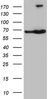
Western blot analysis of extracts (35ug) from 9 different cell lines by using anti-SDCBP monoclonal antibody .

Immunohistochemical staining of paraffin-embedded composit of 3 human ovarian cancer cases using anti-SDCBP 9 mouse monoclonal antibody (BD-PE2721) at 1:100 with Polink2 Broad HRP DAB detection kit; heat-induced epitope retrieval with GBI Citrate pH6.0 HIER buffer using pressure chamber for 3 minutes at 110C. Membrane and cytoplasmic staining is seen in tumor cells with various intensity.

Immunohistochemical staining of paraffin-embedded Carcinoma of Human bladder tissue using anti-SDCBP mouse monoclonal antibody. (BD-PE2721; heat-induced epitope retrieval by 10mM citric buffer, pH6.0, 120°C for 3min)

Immunohistochemical staining of paraffin-embedded mouse lung tissue using anti-SDCBP (SYNTINEN) 9 mouse monoclonal antibody. HIER TEE buffer pH9 at 110C for 10 min, BD-PE2721 (1:100). Detection was done with Klear Mouse (C/N ) DAB Kit.

Immunohistochemical staining of paraffin-embedded human prostate cancer using anti-SDCBP 9 mouse monoclonal antibody (BD-PE2721) at 1:100 with Polink2 Broad HRP DAB detection kit; heat-induced epitope retrieval with GBI Citrate pH6.0 HIER buffer using pressure chamber for 3 minutes at 110C. Membrane and cytoplasmic staining is seen in tumor cells.

Immunohistochemical staining of paraffin-embedded mouse kidney tissue using anti-SDCBP (SYNTINEN) 9 mouse monoclonal antibody. HIER TEE buffer pH9 at 110C for 10 min, BD-PE2721 (1:100). Detection was done with Klear Mouse (C/N ) DAB Kit.

Immunohistochemical staining of paraffin-embedded mouse spleen tissue using anti-SDCBP (SYNTINEN) 9 mouse monoclonal antibody. HIER TEE buffer pH9 at 110C for 10 min, BD-PE2721 (1:100). Detection was done with Klear Mouse (C/N ) DAB Kit.

Western blot analysis of extracts (10ug) from 10 Human tissue by using anti-SDCBP monoclonal antibody at 1:200 (1: Testis; 2: Omentum; 3: Uterus; 4: Breast; 5: Brain; 6: Liver; 7: Ovary; 8: Thyroid gland; 9: colon;10: spleen).

Immunofluorescent staining of MCF-7 cells using anti-SDCBP mouse monoclonal antibody (BD-PE2721, green, 1:50). Actin filaments were labeled with TRITC-phalloidin (red), and nuclear with DAPI (blue).

Immunohistochemical staining of paraffin-embedded Human Kidney tissue using anti-SDCBP mouse monoclonal antibody. (BD-PE2721; heat-induced epitope retrieval by 10mM citric buffer, pH6.0, 120°C for 3min)

相关文献

产品问答

相关产品

免责声明| 法律支持| 联系方式

市场:027-65023363   行政/人事:027-62439686   邮箱:marketing@brainvta.com  

销售总监:张经理  18995532642  华东区:陈经理 18013970337   华南区:王经理 13100653525   华中/西区:杨经理 18186518905   华北区:张经理 18893721749

地址:中国武汉东湖高新区光谷七路128号中科开物产业园1号楼

Copyright © 武汉枢密脑科学技术有限公司. All RIGHTS RESERVED.
鄂ICP备2021009124号 DIGITAL BY VTHINK