Cn|En

现货抗体产品库

PD-1 (PDCD1) (19Q6) Mouse Monoclonal antibody

产品详情 相关文献 产品问答 相关产品

产品基本信息

产品货号
BD-PE2460
产品名称
PD-1 (PDCD1) (19Q6) Mouse Monoclonal antibody
类别
常规抗体
基因名称
PD1
推荐应用
WB
反应种属
Human,Mouse,Dog,Rat
存储缓冲液
PBS (pH 7.3) containing 1% BSA, 50% glycerol and 0.02% New type preservative N.
免疫原
Full length human recombinant protein of human PDCD1 (NP_005009) produced in HEK293T cell.
特异性
Validated through 10k CHIP testing,using a High-Density Protein Microarray that was spotted with ~17,000 various over-expressed proteins to test for antibody specificity.
稀释度
IHC 1:1000, WB 1:500, IF 1:100
预测分子量
29.2kDa
运输及保存条件
-20°C/1 year
宿主
Mouse
同种型
IgG2a
背景介绍
This gene encodes a cell surface membrane protein of the immunoglobulin superfamily. This protein is expressed in pro-B-cells and is thought to play a role in their differentiation. In mice, expression of this gene is induced in the thymus when anti-CD3 antibodies are injected and large numbers of thymocytes undergo apoptosis. Mice deficient for this gene bred on a BALB/c background developed dilated cardiomyopathy and died from congestive heart failure. These studies suggest that this gene product may also be important in T cell function and contribute to the prevention of autoimmune diseases. [provided by RefSeq, Jul
期货
现货
纯化
Purified from mouse ascites fluids or tissue culture supernatant by affinity chromatography (protein A/G)

OriGene overexpression protein microarray chip was immunostained with UltraMAB anti-PDCD1 mouse monoclonal antibody (BD-PE2460). The positive reactive proteins are highlighted with two red arrows in the enlarged subarray. All the positive controls spotted in this subarray are also labeled for clarification (1:100).

Sequential double staining of paraffin human melanoma using b-catenin (red) and PD1 BD-PE2460 (brown). Both abs at 1:800 dilution of 1mg/mL; detection with Polink2 HRP DAB followed by Polink2 Broad AP. Anti-PD1: heat-induced epitope retrieval with Accel; anti-b-catenin: citrate pH6.0. Image shows tumor cells are strongly positve for b-catenin (red) and negative for PD1. The activated T cells (brown) show strong membranous and cytoplasmic staining for PD1 and no staining with b-catenin.

Sequential double staining of paraffin human lung using anti-b-Catenin (red) and anti-PD1 BD-PE2460 (brown). Both antibodies at 1:800 dilution of 1mg/mL. Anti-PD1: heat-induced epitope retrieval with Accel; anti-b-Catenin: citrate pH6.0. Image shows tumor cells are strongly positve for b-catenin (red) and negative for PD1. The arrows point to the activated T cells (brown) showing strong membranous and cytoplasmic staining of PD1 and no staining with b-catenin.

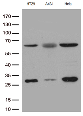
Western blot analysis of extracts (35ug) from PBMCs and PHA-stimulated-PBMCs by using anti-PDCD1 monoclonal antibody (1:250).

Immunohistochemical staining of paraffin-embedded human prostate carcinoma using anti-PD-1 99 mouse monoclonal antibody at 1:800 dilution of 1mg/mL and detection with Polink2 Broad HRP DAB. BD-PE2460 requires heat-induced epitope retrieval with Accel for 3minutes at110C in pressure chamber. The image shows the tumor cells are negative for PD-1 however the activated TCells show strong membranous and cytoplasmic staining.

Immunohistochemical staining of paraffin-embedded human ovarian carcinoma using anti-PD-1 99 mouse monoclonal antibody at 1:800 dilution of 1mg/mL and detection with Polink2 Broad HRP DAB. BD-PE2460 requires heat-induced epitope retrieval with Accel for 3minutes at110C in pressure chamber. The image shows the tumor cells are negative for PD-1 however the activated TCells show strong membranous and cytoplasmic staining.

Immunohistochemical staining of paraffin-embedded human endometrial cancer using anti-PD-1 99 mouse monoclonal antibody at 1:800 dilution of 1mg/mL and detection with Polink2 Broad HRP DAB. BD-PE2460 requires heat-induced epitope retrieval with Accel for 3minutes at110C in pressure chamber. The image shows the tumor cells are negative for PD-1 however the activated TCells show strong membranous and cytoplasmic staining.

Immunohistochemical staining of paraffin-embedded human colon cancer using anti-PD-1 99 mouse monoclonal antibody at 1:800 dilution of 1mg/mL and detection with Polink2 Broad HRP DAB. BD-PE2460 requires heat-induced epitope retrieval with Accel for 3minutes at110C in pressure chamber. The image shows the tumor cells are negative for PD-1 however the activated TCells show strong membranous and cytoplasmic staining.

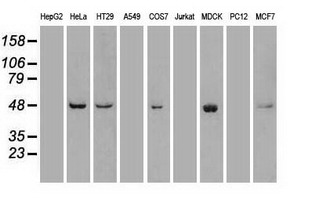
Western blot of cell lysates (35ug) from 9 different cell lines (1: HepG2, 2: HeLa, 3: SV-T2, 4: A549, 5: COS7, 6: Jurkat, 7: MDCK, 8: PC-12, 9: MCF7). Diluation: 1:500.

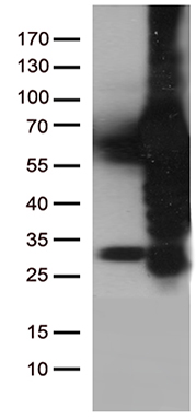
HEK293T cells were transfected with the pCMV6-ENTRY control (Left lane) or pCMV6-ENTRY PDCD1 (Right lane) cDNA for 48 hrs and lysed. Equivalent amounts of cell lysates (5 ug per lane) were separated by SDS-PAGE and immunoblotted with anti-PDCD1 antibody (Cat# BD-PE2460)(1:2000).

Western blot of human tissue lysates (15ug) from 10 different tissues (1: Testis, 2: Omentum, 3: Uterus, 4: Breast, 5: Brain, 6: Thyroid, 7: Colon, 8: Spleen 9: Liver, 10: Ovary). Diluation: 1:500.

Immunocytochemistry staining of PDCD1 -stable-expression cells (left) labeling PDCD1 with mouse anti-PDCD1 monoclonal antibody BD-PE2460 (1:900). The rihgt is negative control.

Immunohistochemical staining of paraffin-embedded human melanoma using anti-PD-1 99 mouse monoclonal antibody at 1:800 dilution of 1mg/mL and detection with Polink2 Broad HRP DAB. BD-PE2460 requires heat-induced epitope retrieval with Accel for 3minutes at110C in pressure chamber. The composite image of 3 melanoma shows the tumor cells are negative for PD-1 however the activated TCells show strong membranous and cytoplasmic staining.

Immunohistochemical staining of 3 paraffin-embedded human lung carcinomas using anti-PD1 99 mouse monoclonal antibody at 1:800 requires HIER Accel [OriGene/GBI Labs in Pressure Chamber for 3 minute on high. Detection of primary antibody was achieved with Polink2 Broad HRP DAB [OriGene/GBI Labs D22].

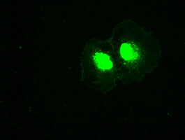
Immunofluorescent staining of PDCD1 -stable-expression cells (left) labeling PDCD1 with mouse monoclonal antibody BD-PE2460 (1:100, green) and nucleus with Hoechst33342 (blue). HEK293T cells serve as negative control (right).

Immunohistochemical staining of paraffin-embedded Human tonsil using anti-PDCD1 mouse monoclonal antibody. (Heat-induced epitope retrieval by 1mM EDTA in 10mM Tris buffer (pH8.5) at 110°C for 10min, BD-PE2460) (1:1200)

相关文献

产品问答

相关产品

免责声明| 法律支持| 联系方式

市场:027-65023363   行政/人事:027-62439686   邮箱:marketing@brainvta.com  

销售总监:张经理  18995532642  华东区:陈经理 18013970337   华南区:王经理 13100653525   华中/西区:杨经理 18186518905   华北区:张经理 18893721749

地址:中国武汉东湖高新区光谷七路128号中科开物产业园1号楼

Copyright © 武汉枢密脑科学技术有限公司. All RIGHTS RESERVED.
鄂ICP备2021009124号 DIGITAL BY VTHINK