PLK1 (17V19) Mouse Monoclonal antibody
产品基本信息
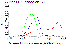
HEK293T cells transfected with either overexpress plasmid (Red), compared to an IgG isotype control, (Green) or empty vector control plasmid (Blue) were immunostained by anti-PLK1 antibody (BD-PE2473), and then analyzed by flow cytometry (1:100).
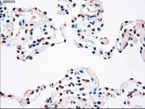
Immunohistochemical staining of paraffin-embedded Human lung tissue within the normal limits using anti-PLK1 mouse monoclonal antibody. (Heat-induced epitope retrieval by 10mM citric buffer, pH6.0, 100°C for 10min, BD-PE2473)
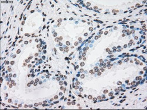
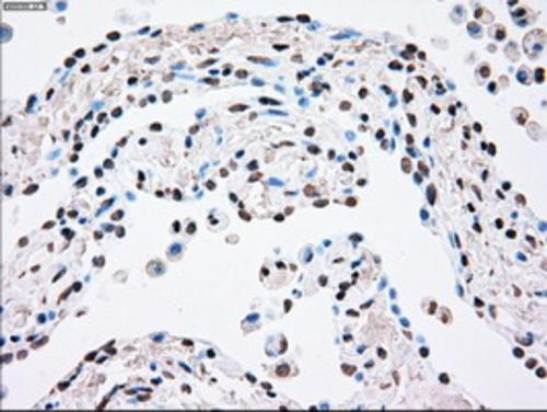
Immunohistochemical staining of paraffin-embedded Human prostate tissue within the normal limits using anti-PLK1 mouse monoclonal antibody. (Heat-induced epitope retrieval by 10mM citric buffer, pH6.0, 100°C for 10min, BD-PE2473)
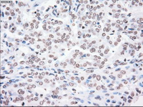
Immunohistochemical staining of paraffin-embedded Adenocarcinoma of Human endometrium tissue using anti-PLK1 mouse monoclonal antibody. (Heat-induced epitope retrieval by 10mM citric buffer, pH6.0, 100°C for 10min, BD-PE2473)
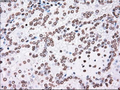
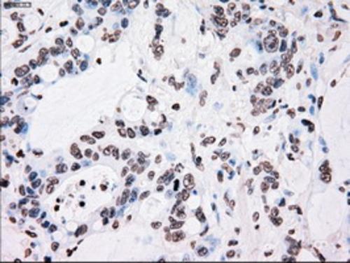
Immunohistochemical staining of paraffin-embedded Carcinoma of Human kidney tissue using anti-PLK1 mouse monoclonal antibody. (Heat-induced epitope retrieval by 10mM citric buffer, pH6.0, 100°C for 10min, BD-PE2473)
Immunohistochemical staining of paraffin-embedded Human endometrium tissue within the normal limits using anti-PLK1 mouse monoclonal antibody. (Heat-induced epitope retrieval by 10mM citric buffer, pH6.0, 100°C for 10min, BD-PE2473)
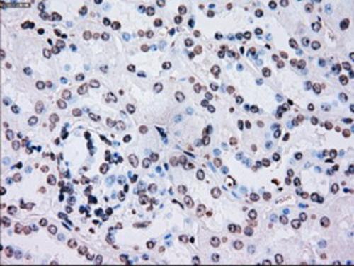
Immunohistochemical staining of paraffin-embedded Human Kidney tissue within the normal limits using anti-PLK1 mouse monoclonal antibody. (Heat-induced epitope retrieval by 10mM citric buffer, pH6.0, 100°C for 10min, BD-PE2473)
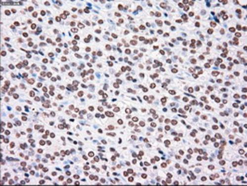
Immunohistochemical staining of paraffin-embedded Carcinoma of Human thyroid tissue using anti-PLK1 mouse monoclonal antibody. (Heat-induced epitope retrieval by 10mM citric buffer, pH6.0, 100°C for 10min, BD-PE2473)
Immunohistochemical staining of paraffin-embedded Adenocarcinoma of Human colon tissue using anti-PLK1 mouse monoclonal antibody. (Heat-induced epitope retrieval by 10mM citric buffer, pH6.0, 100°C for 10min, BD-PE2473)
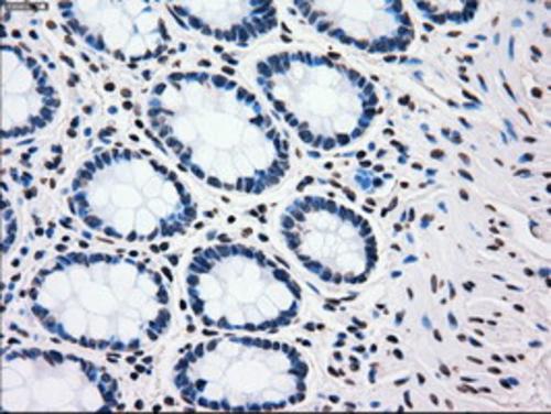
Immunohistochemical staining of paraffin-embedded Human colon tissue within the normal limits using anti-PLK1 mouse monoclonal antibody. (Heat-induced epitope retrieval by 10mM citric buffer, pH6.0, 100°C for 10min, BD-PE2473)
Immunohistochemical staining of paraffin-embedded Carcinoma of Human pancreas tissue using anti-PLK1 mouse monoclonal antibody. (Heat-induced epitope retrieval by 10mM citric buffer, pH6.0, 100°C for 10min, BD-PE2473)
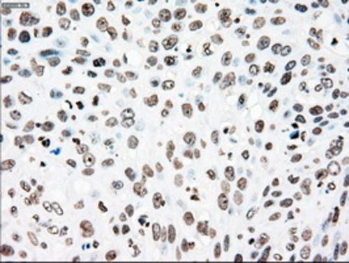
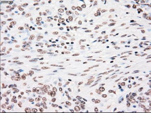
Immunohistochemical staining of paraffin-embedded Adenocarcinoma of Human breast tissue using anti-PLK1 mouse monoclonal antibody. (Heat-induced epitope retrieval by 10mM citric buffer, pH6.0, 100°C for 10min, BD-PE2473)
Immunohistochemical staining of paraffin-embedded Human pancreas tissue within the normal limits using anti-PLK1 mouse monoclonal antibody. (Heat-induced epitope retrieval by 10mM citric buffer, pH6.0, 100°C for 10min, BD-PE2473)
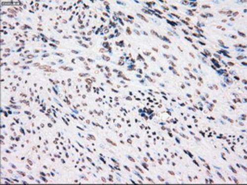
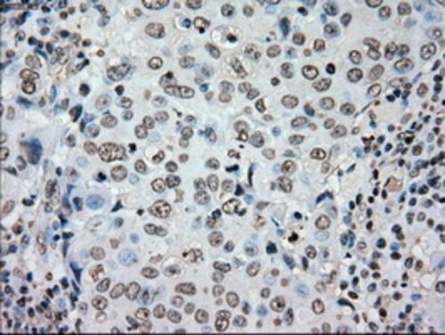
Immunohistochemical staining of paraffin-embedded Carcinoma of Human bladder tissue using anti-PLK1 mouse monoclonal antibody. (Heat-induced epitope retrieval by 10mM citric buffer, pH6.0, 100°C for 10min, BD-PE2473)
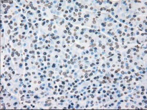
Immunohistochemical staining of paraffin-embedded Carcinoma of Human lung tissue using anti-PLK1 mouse monoclonal antibody. (Heat-induced epitope retrieval by 10mM citric buffer, pH6.0, 100°C for 10min, BD-PE2473)
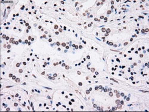
Immunohistochemical staining of paraffin-embedded Carcinoma of Human prostate tissue using anti-PLK1 mouse monoclonal antibody. (Heat-induced epitope retrieval by 10mM citric buffer, pH6.0, 100°C for 10min, BD-PE2473)
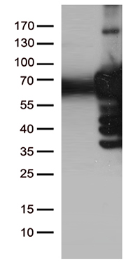
HEK293T cells were transfected with the pCMV6-ENTRY control (Left lane) or pCMV6-ENTRY PLK1 (Right lane) cDNA for 48 hrs and lysed. Equivalent amounts of cell lysates (5 ug per lane) were separated by SDS-PAGE and immunoblotted with anti-PLK1 (Cat# BD-PE2473). Positive lysates (100ug) and (20ug) can be purchased separately from OriGene.
相关文献
产品问答
相关产品

市场:027-65023363 行政/人事:027-62439686 邮箱:marketing@brainvta.com 客服:18140661572(活动咨询、售后反馈等)
销售总监:张经理 18995532642 华东区:陈经理 18013970337 华南区:王经理 13100653525 华中/西区:杨经理 18186518905 华北区:张经理 18893721749
地址:中国武汉东湖高新区光谷七路128号中科开物产业园1号楼
Copyright © 武汉枢密脑科学技术有限公司. All RIGHTS RESERVED.
鄂ICP备2021009124号 DIGITAL BY VTHINK
