GATA3 (18Q10) Mouse Monoclonal antibody
产品基本信息
OriGene overexpression protein microarray chip was immunostained with UltraMAB anti-GATA3 mouse monoclonal antibody (BD-PE0583). The positive reactive proteins are highlighted with two red arrows in the enlarged subarray. All the positive controls spotted in this subarray are also labeled for clarification (1:100).
Immunohistochemical staining of paraffin-embedded human urothelial carcinoma using anti-GATA3 mouse monoclonal antibody. HIER pretreatment was done with 1mM EDTA in 10mM Tris buffer (pH8.0) at 120°C for 2.5 minutes. BD-PE0583 was diluted 1:50 and detection was done with HRP secondary and DAB chromogen. Strong nuclear stain is seen in the tumor cells.
Immunohistochemical staining of paraffin-embedded human bladder cancer using anti-GATA3 at 1:200 dilution of 1 mg/mL and detection with Polink2 Broad HRP DAB. BD-PE0583 requires heat-induced epitope retrieval with ACCEL (pH8.7) biocare pressure cooker at 110°C for 3minutes. The image shows nuclear staining in tumor cells.
Immunohistochemical staining of paraffin-embedded human kidney tissue within the normal limits using anti-GATA3 mouse monoclonal antibody. HIER pretreatment was done with 1mM EDTA in 10mM Tris buffer (pH8.0) at 120°C for 2.5 minutes. BD-PE0583 was diluted 1:50, detection was done with HRP secondary and DAB chromogen. Weak nuclear stain is seen in the tubule epithelial cells of the kidney.
HEK293T cells were transfected with the pCMV6-ENTRY control (Left lane) or pCMV6-ENTRY GATA3 (Right lane) cDNA for 48 hrs and lysed. Equivalent amounts of cell lysates (5 ug per lane) were separated by SDS-PAGE and immunoblotted with anti-GATA3 (1:500).
Immunocytochemistry staining of MCF-7 cells using anti-GATA3 mouse monoclonal antibody (BD-PE0583) (Left). The right is negative control. (1:2000)
Immunohistochemical staining of paraffin-embedded carcinoma of human prostate tissue using anti-GATA3 mouse monoclonal antibody. HIER pretreatment was done with 1mM EDTA in 10mM Tris buffer (pH8.0) at 120°C for 2.5 minutes. BD-PE0583 was diluted 1:50 and detection was done with HRP secondary and DAB chromogen. Stong nuclear stain was seen in the tumor cells.
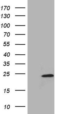
Western blot analysis of extracts (35ug) from 2 different cell by using anti-GATA3 monoclonal antibody (1:500).
Immunohistochemical staining of paraffin-embedded at margins of adenocarcinoma of human breast tissue using anti-GATA3 mouse monoclonal antibody. HIER pretreatment was done with 1mM EDTA in 10mM Tris buffer (pH8.0) at 120°C for 2.5 minutes. BD-PE0583 was diluted 1:50 and detection was done with HRP secondary and DAB chromogen. Strong nuclear stain is seen in the epithelial cells.
相关文献
产品问答
相关产品
市场:027-65023363 行政/人事:027-62439686 邮箱:marketing@brainvta.com
销售总监:张经理 18995532642 华东区:陈经理 18013970337 华南区:王经理 13100653525 华中/西区:杨经理 18186518905 华北区:张经理 18893721749
地址:中国武汉东湖高新区光谷七路128号中科开物产业园1号楼
Copyright © 武汉枢密脑科学技术有限公司. All RIGHTS RESERVED.
鄂ICP备2021009124号 DIGITAL BY VTHINK