PON1 (12V14) Mouse Monoclonal antibody
产品基本信息
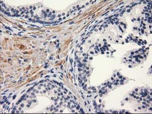
Immunohistochemical staining of paraffin-embedded Human prostate tissue within the normal limits using anti-PON1 mouse monoclonal antibody. (Heat-induced epitope retrieval by 10mM citric buffer, pH6.0, 100°C for 10min, BD-PE0286)
Immunohistochemical staining of paraffin-embedded Human endometrium tissue within the normal limits using anti-PON1 mouse monoclonal antibody. (Heat-induced epitope retrieval by 10mM citric buffer, pH6.0, 100°C for 10min, BD-PE0286)
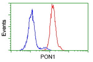
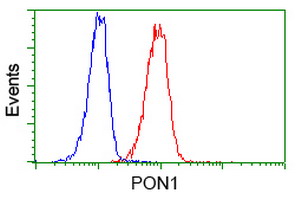
HEK293T cells transfected with either overexpress plasmid (Red) or empty vector control plasmid (Blue) were immunostained by anti-PON1 antibody (BD-PE0286), and then analyzed by flow cytometry.
Flow cytometric Analysis of Hela cells, using anti-PON1 antibody (BD-PE0286), (Red), compared to a nonspecific negative control antibody , (Blue).
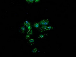
Anti-PON1 mouse monoclonal antibody (BD-PE0286) immunofluorescent staining of COS7 cells transiently transfected by pCMV6-ENTRY PON1 .
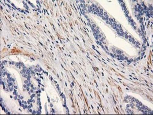
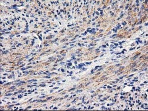
Immunohistochemical staining of paraffin-embedded Carcinoma of Human prostate tissue using anti-PON1 mouse monoclonal antibody. (Heat-induced epitope retrieval by 10mM citric buffer, pH6.0, 100°C for 10min, BD-PE0286)
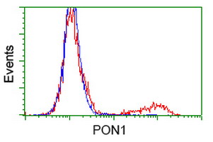
Flow cytometric Analysis of Jurkat cells, using anti-PON1 antibody (BD-PE0286), (Red), compared to a nonspecific negative control antibody , (Blue).
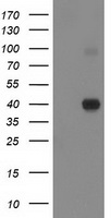
HEK293T cells were transfected with the pCMV6-ENTRY control (Left lane) or pCMV6-ENTRY PON1 (Right lane) cDNA for 48 hrs and lysed. Equivalent amounts of cell lysates (5 ug per lane) were separated by SDS-PAGE and immunoblotted with anti-PON1 (BD-PE0286). Positive lysates (100ug) and (20ug) can be purchased separately from OriGene.
相关文献
产品问答
相关产品

市场:027-65023363 行政/人事:027-62439686 邮箱:marketing@brainvta.com 客服:18140661572(活动咨询、售后反馈等)
销售总监:张经理 18995532642 华东区:陈经理 18013970337 华南区:王经理 13100653525 华中/西区:杨经理 18186518905 华北区:张经理 18893721749
地址:中国武汉东湖高新区光谷七路128号中科开物产业园1号楼
Copyright © 武汉枢密脑科学技术有限公司. All RIGHTS RESERVED.
鄂ICP备2021009124号 DIGITAL BY VTHINK
