产品名称
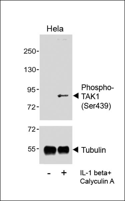
Phospho-TAK1 (Ser439) Rabbit Polyclonal Antibody
别名
Mitogen-activated protein kinase kinase kinase 7, 2.7.11.25, Transforming growth factor-beta-activated kinase 1, TGF-beta-activated kinase 1, MAP3K7, TAK1
存储缓冲液
Purified polyclonal antibody supplied in PBS with 0.09% (W/V) New?type?preservative?N. This antibody is purified through a protein A column, followed by peptide affinity purification.
Human Swissprot No.
O43318
特异性
This Phospho-TAK1 (Ser439) antibody is generated from a rabbit immunized with a KLH conjugated synthetic peptide between 410-444 amino acids from the human TAK1.
运输及保存条件
Maintain refrigerated at 2-8°C for up to 2 weeks. For long term storage store at -20°C in small aliquots to prevent freeze-thaw cycles.
背景介绍
Serine/threonine kinase which acts as an essential component of the MAP kinase signal transduction pathway. Plays an important role in the cascades of cellular responses evoked by changes in the environment. Mediates signal transduction of TRAF6, various cytokines including interleukin-1 (IL-1), transforming growth factor-beta (TGFB), TGFB-related factors like BMP2 and BMP4, toll-like receptors (TLR), tumor necrosis factor receptor CD40 and B-cell receptor (BCR). Ceramides are also able to activate MAP3K7/TAK1. Once activated, acts as an upstream activator of the MKK/JNK signal transduction cascade and the p38 MAPK signal transduction cascade through the phosphorylation and activation of several MAP kinase kinases like MAP2K1/MEK1, MAP2K3/MKK3, MAP2K6/MKK6 and MAP2K7/MKK7. These MAP2Ks in turn activate p38 MAPKs, c-jun N-terminal kinases (JNKs) and I-kappa-B kinase complex (IKK). Both p38 MAPK and JNK pathways control the transcription factors activator protein-1 (AP-1), while nuclear factor-kappa B is activated by IKK. MAP3K7 activates also IKBKB and MAPK8/JNK1 in response to TRAF6 signaling and mediates BMP2- induced apoptosis. In osmotic stress signaling, plays a major role in the activation of MAPK8/JNK1, but not that of NF-kappa-B. Promotes TRIM5 capsid-specific restriction activity.
组织表达
Isoform 1A is the most abundant in ovary, skeletal muscle, spleen and blood mononuclear cells. Isoform 1B is highly expressed in brain, kidney and small intestine. Isoform 1C is the major form in prostate. Isoform 1D is the less abundant form
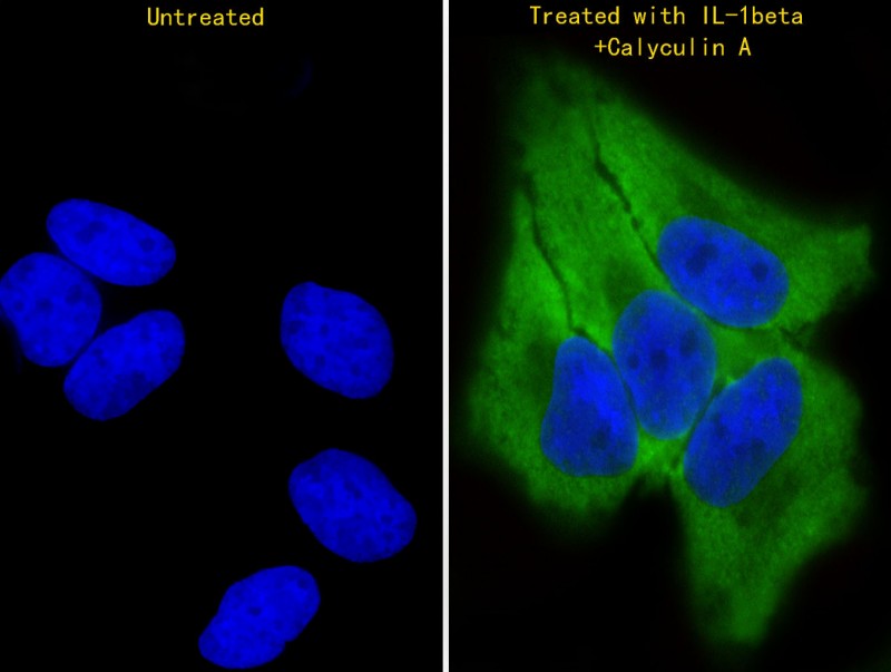
细胞定位
Cytoplasm. Cell membrane; Peripheral membrane protein; Cytoplasmic side. Note=Although the majority of MAP3K7/TAK1 is found in the cytosol, when complexed with TAB1/MAP3K7IP1 and TAB2/MAP3K7IP2, it is also localized at the cell membrane
功能
Serine/threonine kinase which acts as an essential component of the MAP kinase signal transduction pathway. Plays an important role in the cascades of cellular responses evoked by changes in the environment. Mediates signal transduction of TRAF6, various cytokines including interleukin-1 (IL-1), transforming growth factor-beta (TGFB), TGFB-related factors like BMP2 and BMP4, toll-like receptors (TLR), tumor necrosis factor receptor CD40 and B-cell receptor (BCR). Ceramides are also able to activate MAP3K7/TAK1. Once activated, acts as an upstream activator of the MKK/JNK signal transduction cascade and the p38 MAPK signal transduction cascade through the phosphorylation and activation of several MAP kinase kinases like MAP2K1/MEK1, MAP2K3/MKK3, MAP2K6/MKK6 and MAP2K7/MKK7. These MAP2Ks in turn activate p38 MAPKs, c-jun N-terminal kinases (JNKs) and I-kappa-B kinase complex (IKK). Both p38 MAPK and JNK pathways control the transcription factors activator protein-1 (AP-1), while nuclear factor-kappa B is activated by IKK. MAP3K7 activates also IKBKB and MAPK8/JNK1 in response to TRAF6 signaling and mediates BMP2-induced apoptosis. In osmotic stress signaling, plays a major role in the activation of MAPK8/JNK1, but not that of NF-kappa-B. Promotes TRIM5 capsid-specific restriction activity. Phosphorylates RIPK1 at 'Ser-321' which positively regulates RIPK1 interaction with RIPK3 to promote necroptosis but negatively regulates RIPK1 kinase activity and its interaction with FADD to mediate apoptosis (By similarity).