Cn|En

现货抗体产品库

FGFR1 Rabbit Polyclonal Antibody (Center)

产品详情 相关文献 产品问答 相关产品

产品基本信息

产品货号
BD-PB4898
产品名称
FGFR1 Rabbit Polyclonal Antibody (Center)
别名
Fibroblast growth factor receptor 1, FGFR-1, Basic fibroblast growth factor receptor 1, BFGFR, bFGF-R-1, Fms-like tyrosine kinase 2, FLT-2, N-sam, Proto-oncogene c-Fgr, CD331, FGFR1, BFGFR, CEK, FGFBR, FLG, FLT2, HBGFR
类别
常规抗体
推荐应用
WB
反应种属
Human, Rat, Mouse
存储缓冲液
Purified polyclonal antibody supplied in PBS with 0.09% (W/V) New type preservative N. This antibody is purified through a protein A column, followed by peptide affinity purification.
Human Swissprot No.
P11362
特异性
This FGFR1 antibody is generated from a rabbit immunized with a KLH conjugated synthetic peptide between 291-324 amino acids from the Central region of human FGFR1.
稀释度
FC~~1:25;WB~~1:1000
预测分子量
92kDa
运输及保存条件
Maintain refrigerated at 2-8°C for up to 2 weeks. For long term storage store at -20°C in small aliquots to prevent freeze-thaw cycles.
宿主
Rabbit
同种型
Rabbit IgG
背景介绍
Tyrosine-protein kinase that acts as cell-surface receptor for fibroblast growth factors and plays an essential role in the regulation of embryonic development, cell proliferation, differentiation and migration. Required for normal mesoderm patterning and correct axial organization during embryonic development, normal skeletogenesis and normal development of the gonadotropin-releasing hormone (GnRH) neuronal system. Phosphorylates PLCG1, FRS2, GAB1 and SHB. Ligand binding leads to the activation of several signaling cascades. Activation of PLCG1 leads to the production of the cellular signaling molecules diacylglycerol and inositol 1,4,5-trisphosphate. Phosphorylation of FRS2 triggers recruitment of GRB2, GAB1, PIK3R1 and SOS1, and mediates activation of RAS, MAPK1/ERK2, MAPK3/ERK1 and the MAP kinase signaling pathway, as well as of the AKT1 signaling pathway. Promotes phosphorylation of SHC1, STAT1 and PTPN11/SHP2. In the nucleus, enhances RPS6KA1 and CREB1 activity and contributes to the regulation of transcription. FGFR1 signaling is down-regulated by IL17RD/SEF, and by FGFR1 ubiquitination, internalization and degradation.
组织表达
Detected in astrocytoma, neuroblastoma and adrenal cortex cell lines. Some isoforms are detected in foreskin fibroblast cell lines, however isoform 17, isoform 18 and isoform 19 are not detected in these cells.
细胞定位
Cell membrane; Single-pass type I membrane protein. Nucleus. Cytoplasm, cytosol. Cytoplasmic vesicle. Note=After ligand binding, both receptor and ligand are rapidly internalized. Can translocate to the nucleus after internalization, or by translocation from the endoplasmic reticulum or Golgi apparatus to the cytosol, and from there to the nucleus
功能
Tyrosine-protein kinase that acts as cell-surface receptor for fibroblast growth factors and plays an essential role in the regulation of embryonic development, cell proliferation, differentiation and migration. Required for normal mesoderm patterning and correct axial organization during embryonic development, normal skeletogenesis and normal development of the gonadotropin-releasing hormone (GnRH) neuronal system. Phosphorylates PLCG1, FRS2, GAB1 and SHB. Ligand binding leads to the activation of several signaling cascades. Activation of PLCG1 leads to the production of the cellular signaling molecules diacylglycerol and inositol 1,4,5-trisphosphate. Phosphorylation of FRS2 triggers recruitment of GRB2, GAB1, PIK3R1 and SOS1, and mediates activation of RAS, MAPK1/ERK2, MAPK3/ERK1 and the MAP kinase signaling pathway, as well as of the AKT1 signaling pathway. Promotes phosphorylation of SHC1, STAT1 and PTPN11/SHP2. In the nucleus, enhances RPS6KA1 and CREB1 activity and contributes to the regulation of transcription. FGFR1 signaling is down-regulated by IL17RD/SEF, and by FGFR1 ubiquitination, internalization and degradation.
期货
现货

Flow cytometric analysis of SH-SY5Y cells using FGFR1 Antibody (Center)(green) compared to an isotype control of rabbit IgG(blue). BD-PB4898 was diluted at 1:25 dilution. An Alexa Fluor® 488 goat anti-rabbit lgG at 1:400 dilution was used as the secondary antibody.

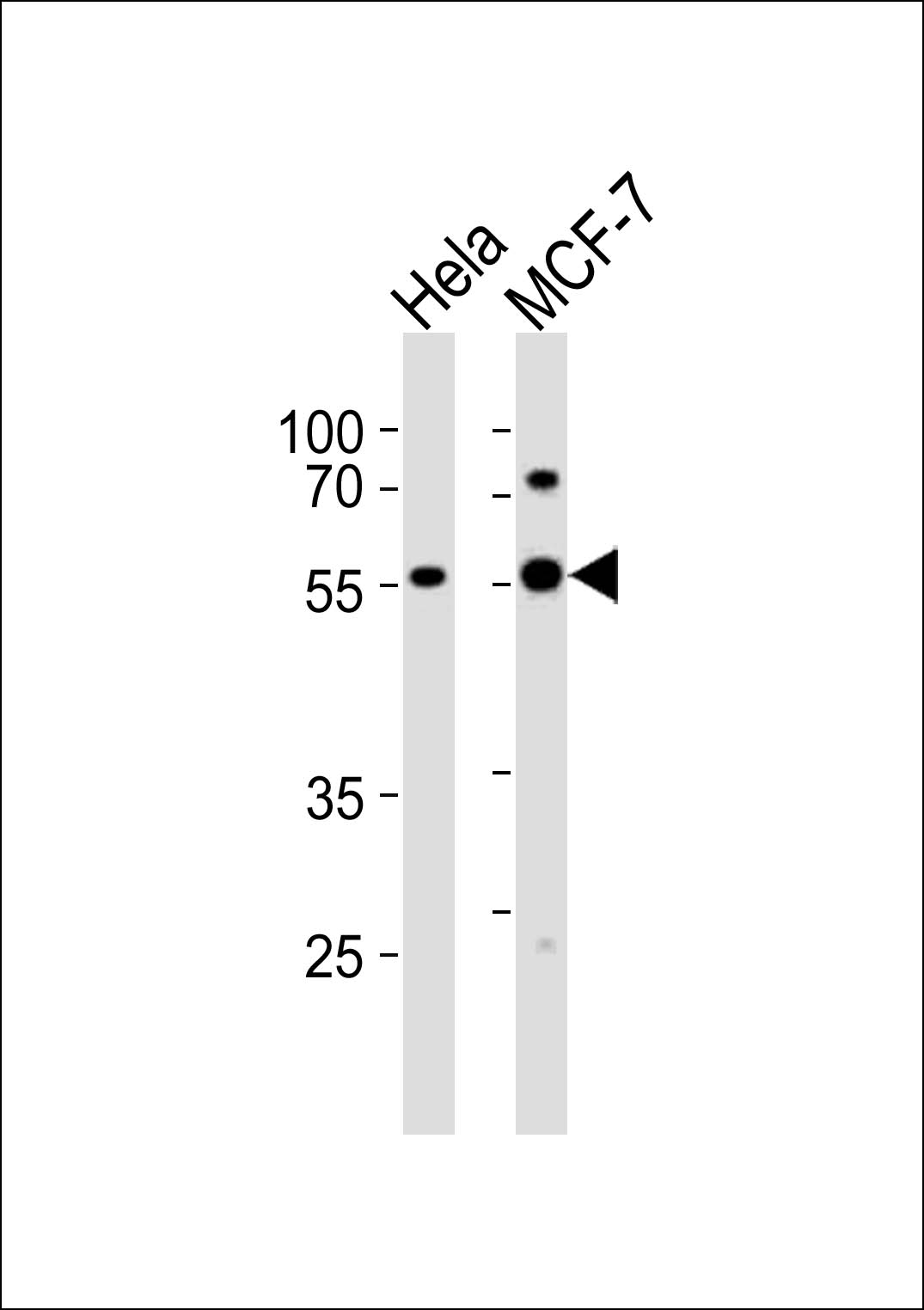
Western blot analysis of lysates from Hela, HepG2, MCF-7, SH-SY5Y, mouse NIH/3T3 cell line (from left to right), using FGFR1 Antibody (Center)at 1:10000 dilution was used as the secondary antibody. Lysates at 20ug per lane.

相关文献

产品问答

相关产品

免责声明| 法律支持| 联系方式

市场:027-65023363   行政/人事:027-62439686   邮箱:marketing@brainvta.com  

销售总监:张经理  18995532642  华东区:陈经理 18013970337   华南区:王经理 13100653525   华中/西区:杨经理 18186518905   华北区:张经理 18893721749

地址:中国武汉东湖高新区光谷七路128号中科开物产业园1号楼

Copyright © 武汉枢密脑科学技术有限公司. All RIGHTS RESERVED.
鄂ICP备2021009124号 DIGITAL BY VTHINK