CAV2 Rabbit Polyclonal Antibody (N-term)
产品基本信息
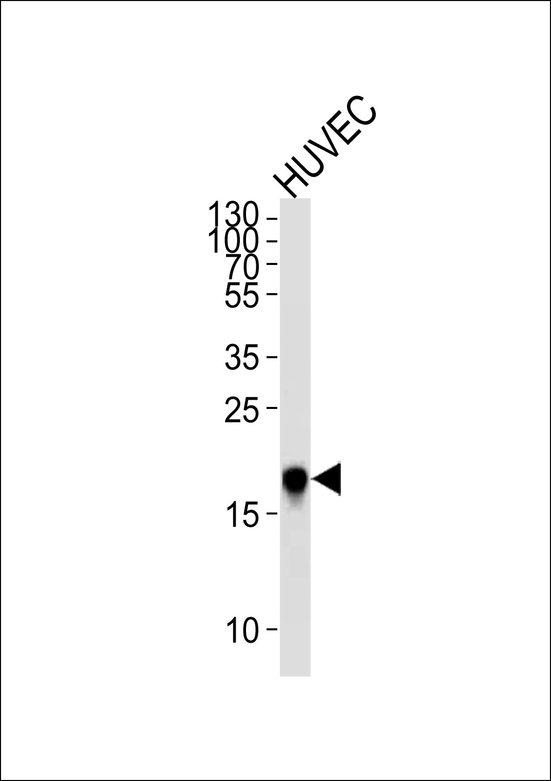
Western blot analysis of lysate from HUVEC cell line,using CAV2 Antibody (N-term)at 1:5000 dilution was used as the secondary antibody.Lysate at 35ug per lane.
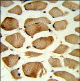
Formalin-fixed and paraffin-embedded human skeletal muscle reacted with CAV2 Antibody (N-term), which was peroxidase-conjugated to the secondary antibody, followed by DAB staining. This data demonstrates the use of this antibody for immunohistochemistry; clinical relevance has not been evaluated.
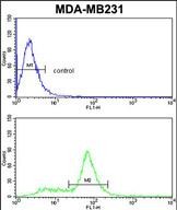
CAV2 Antibody (N-term) flow cytometric analysis of MDA-MB231 cells (bottom histogram) compared to a negative control cell (top histogram).FITC-conjugated goat-anti-rabbit secondary antibodies were used for the analysis.
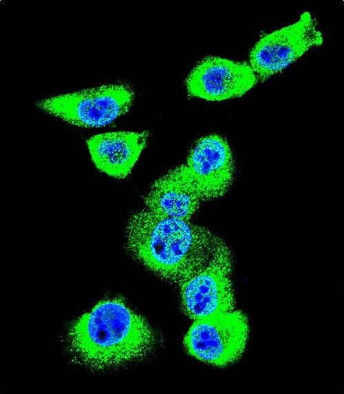
Confocal immunofluorescent analysis of CAV2 Antibody (N-term)with MDA-MB231 cell followed by Alexa Fluor 488-conjugated goat anti-rabbit lgG (green). DAPI was used to stain the cell nuclear (blue).
相关文献
产品问答
相关产品

市场:027-65023363 行政/人事:027-62439686 邮箱:marketing@brainvta.com 客服:18140661572(活动咨询、售后反馈等)
销售总监:张经理 18995532642 华东区:陈经理 18013970337 华南区:王经理 13100653525 华中/西区:杨经理 18186518905 华北区:张经理 18893721749
地址:中国武汉东湖高新区光谷七路128号中科开物产业园1号楼
Copyright © 武汉枢密脑科学技术有限公司. All RIGHTS RESERVED.
鄂ICP备2021009124号 DIGITAL BY VTHINK
